. 2022 Nov-Dec:128:1-12.
doi: 10.1016/j.diff.2022.09.002. Epub 2022 Sep 24.

Myhre syndrome is caused by dominant-negative dysregulation of SMAD4 and other co-factors

Affiliations

Myhre syndrome is caused by dominant-negative dysregulation of SMAD4 and other co-factors

Dimuthu Alankarage et al. Differentiation. 2022 Nov-Dec.

Abstract

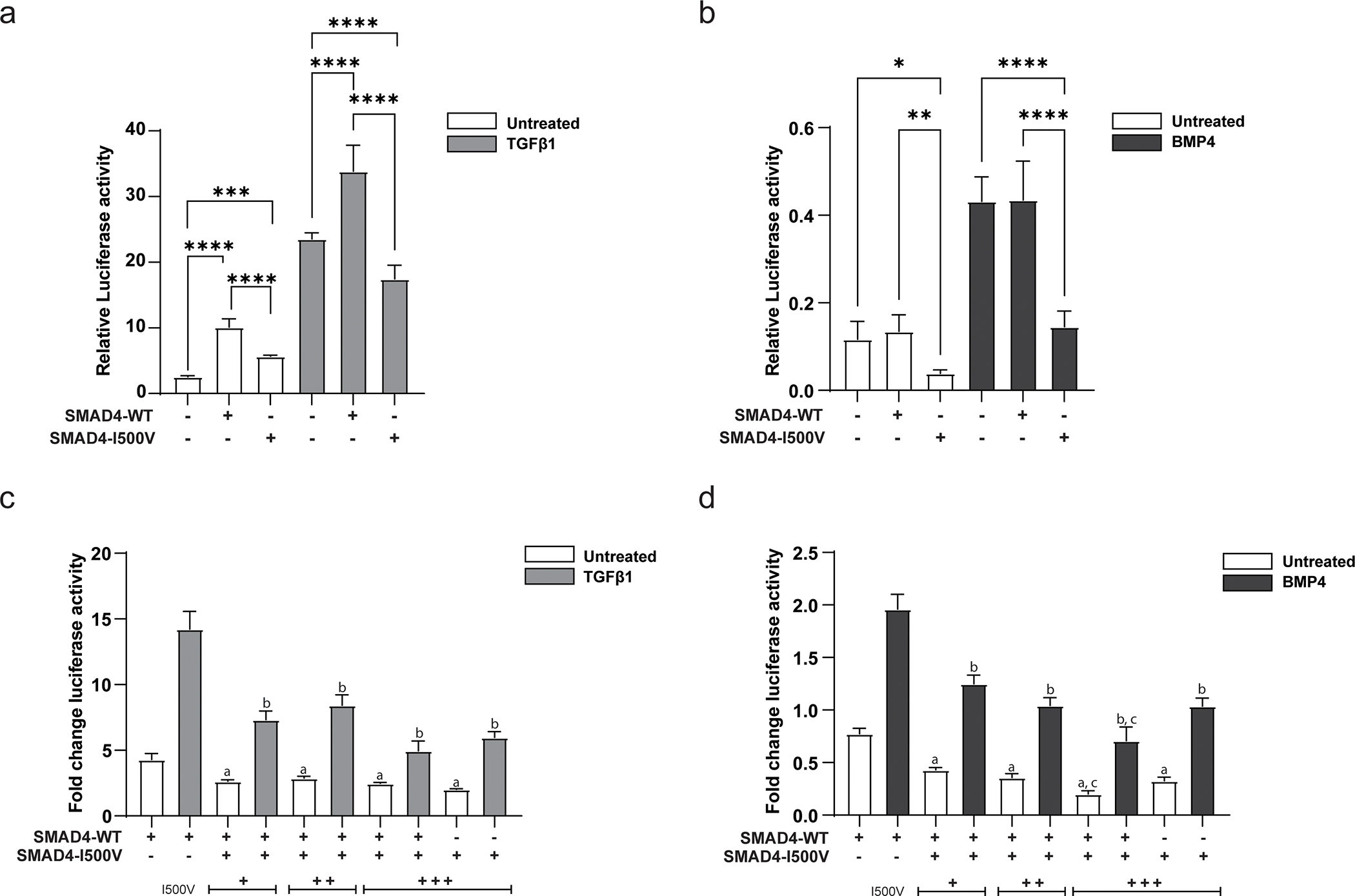
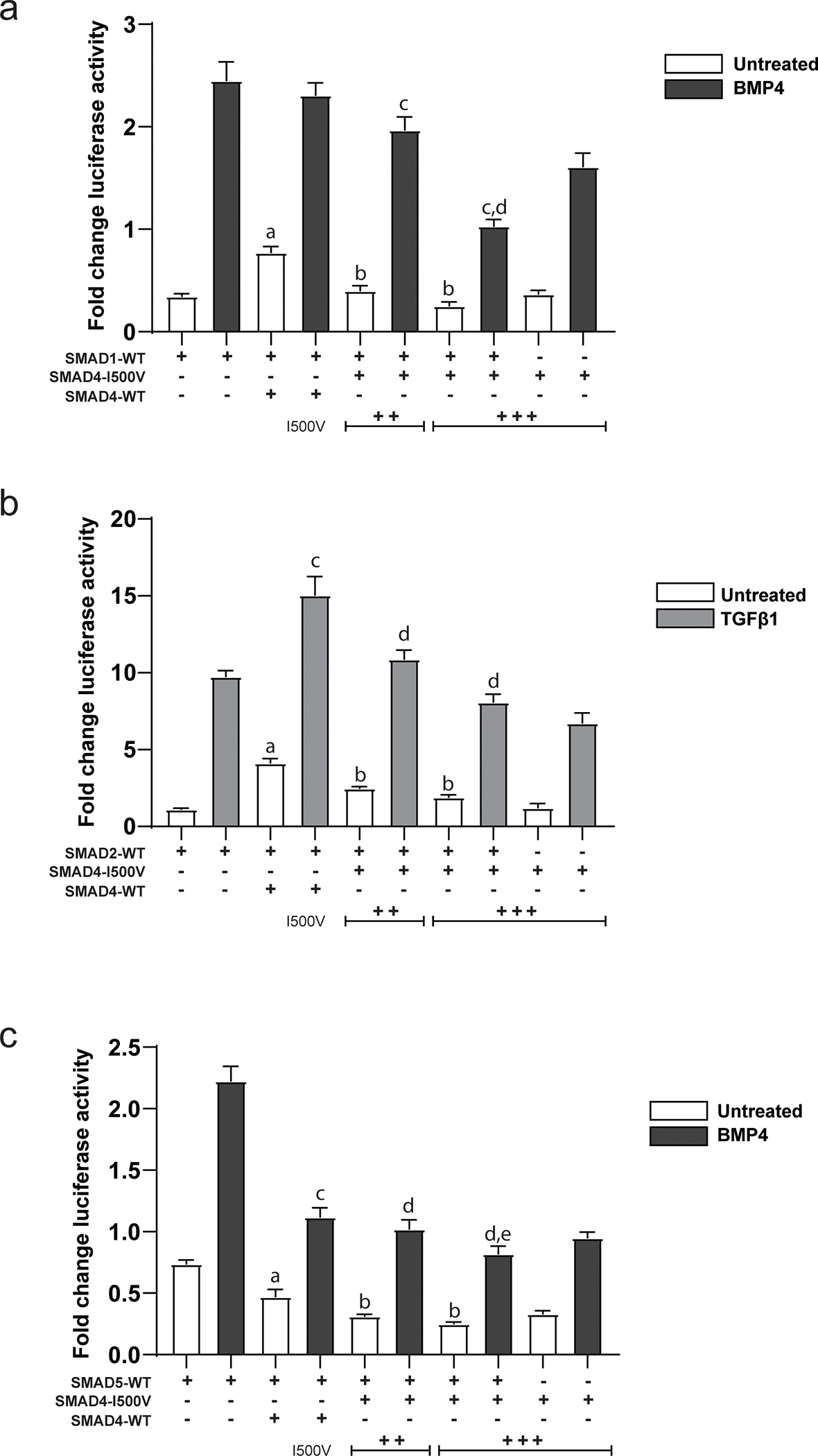
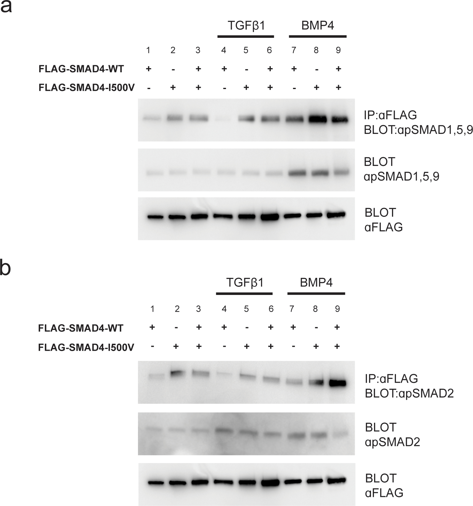
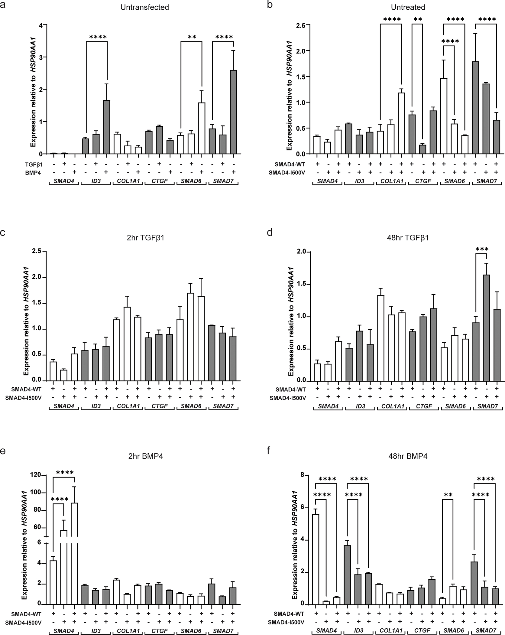
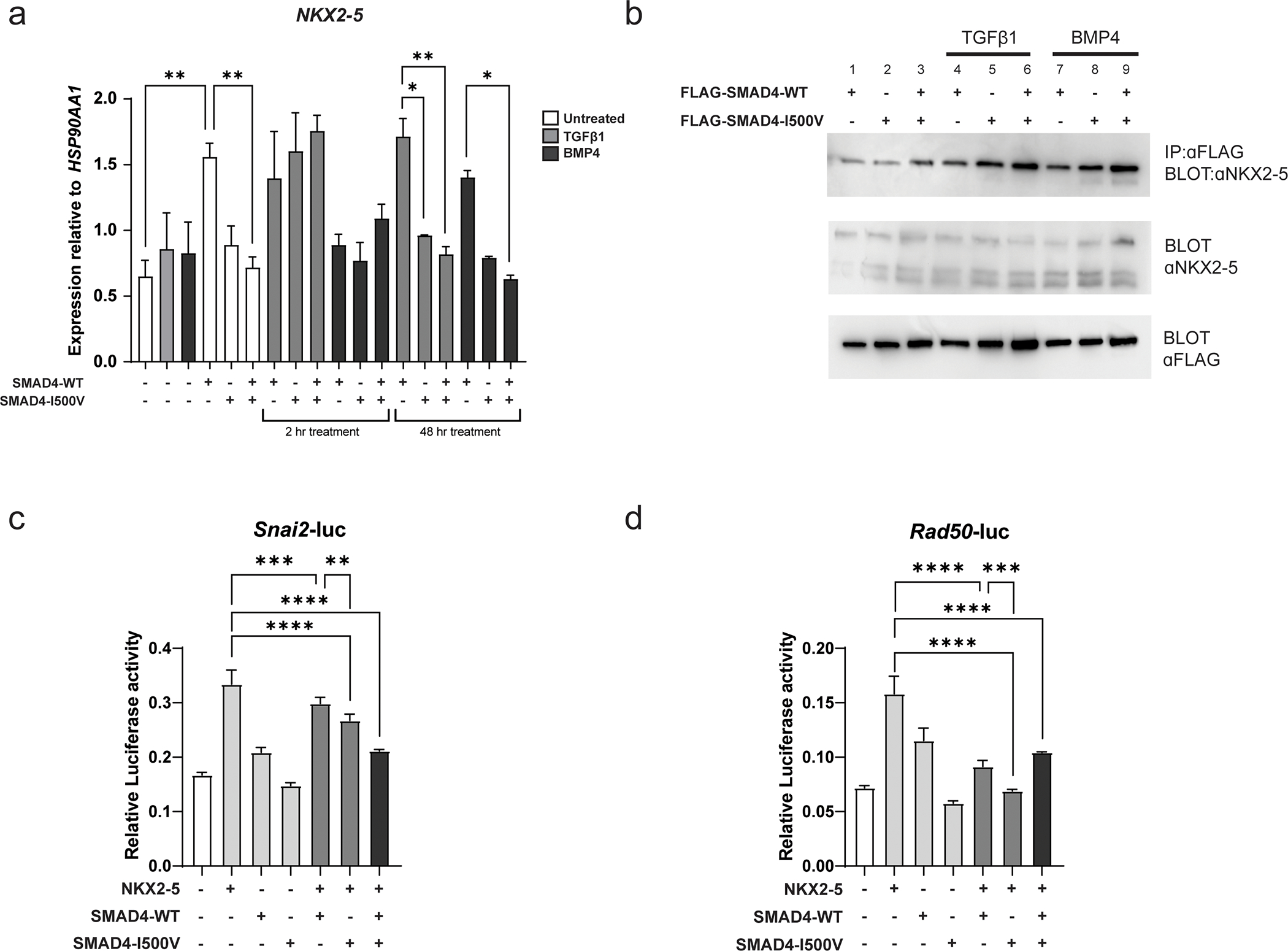
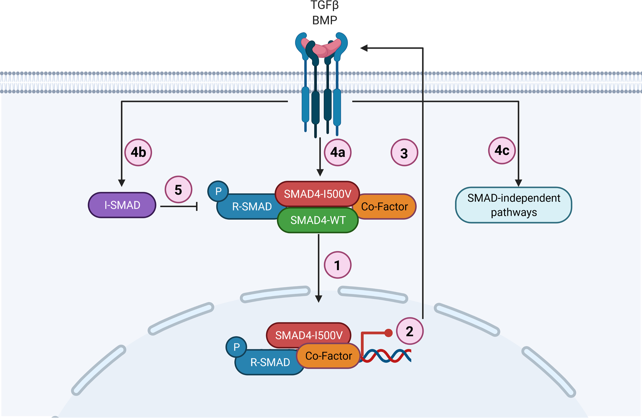
Myhre syndrome is a connective tissue disorder characterized by congenital cardiovascular, craniofacial, respiratory, skeletal, and cutaneous anomalies as well as intellectual disability and progressive fibrosis. It is caused by germline variants in the transcriptional co-regulator SMAD4 that localize at two positions within the SMAD4 protein, I500 and R496, with I500 V/T/M variants more commonly identified in individuals with Myhre syndrome. Here we assess the functional impact of SMAD4-I500V variant, identified in two previously unpublished individuals with Myhre syndrome, and provide novel insights into the molecular mechanism of SMAD4-I500V dysfunction. We show that SMAD4-I500V can dimerize, but its transcriptional activity is severely compromised. Our data show that SMAD4-I500V acts dominant-negatively on SMAD4 and on receptor-regulated SMADs, affecting transcription of target genes. Furthermore, SMAD4-I500V impacts the transcription and function of crucial developmental transcription regulator, NKX2-5. Overall, our data reveal a dominant-negative model of disease for SMAD4-I500V where the function of SMAD4 encoded on the remaining allele, and of co-factors, are perturbed by the continued heterodimerization of the variant, leading to dysregulation of TGF and BMP signaling. Our findings not only provide novel insights into the mechanism of Myhre syndrome pathogenesis but also extend the current knowledge of how pathogenic variants in SMAD proteins cause disease.

Keywords: Dominant-negative; Myhre syndrome; NKX2-5; SMAD4; TGF/BMP signaling; Transcriptional regulation.


Conflict of interest statement

Declaration of competing interest None.

Figures

Fig. 1.
Fig. 1.. SMAD4-I500V shows greatly reduced ability to activate target promoters and acts dominant-negatively on WT-SMAD4.
(A) Impact of the I500V variant on basal and TGFβ1-activated SBE(4)-luc was assessed by luciferase assays, ***<0.001, **** < 0.0001, n = 9–12. (B) Impact of the I500V variant on basal and BMP4-activated Xvent2-luc was assessed by luciferase assays, * < 0.05, ** < 0.01, **** < 0.0001, n = 9–12. Luciferase activity was normalized to Renilla and analyzed by one-way ANOVA, presented as mean ± SD. (C) Dominant-negative response of the I500V variant on WT-SMAD4 action on SBE(4)-luc in basal and TGFβ1-treated cells. (D) Dominant-negative response of the I500V variant on WT-SMAD4 action on Xvent2-luc in basal and BMP4-treated cells, a: comparison between WT-SMAD4- and WT/I500V- or I500V-transfected cells in basal conditions, p < 0.0001; b: comparison between WT-SMAD4- and WT/I500V- or I500V-transfected cells in TGFβ1/BMP4-treated cells, p < 0.001–0.0001; c: comparison between WT/I500V- and I500V-transfected cells in basal and BMP4-treated cells, p < 0.001–0.01. Luciferase activity was normalized to vector transfected cells within each treatment group, analyzed by one-way ANOVA, presented as mean ± SD, n = 6.
Fig. 2.
Fig. 2.. Dominant-negative activity of SMAD4-I500V inhibits function of R-SMADs.
(A) Action of the I500V variant on SMAD1 activity on the Xvent2-luc reporter in basal and BMP4-treated cells was assessed, a: comparison between SMAD1- and SMAD1/WT-SMAD4- transfected cells in basal conditions, p < 0.0001; b: comparison between SMAD1/WT-SMAD4- and SMAD1/SMAD4-I500V- transfected cells in basal conditions, p < 0.0001; c: comparison between SMAD1/WT-SMAD4- and SMAD1/SMAD4-I500V- transfected cells in BMP4-treated cells, p < 0.0001; d: comparison between SMAD4-I500V- and SMAD1/SMAD4-I500V- transfected cells in BMP4-treated cells, p < 0.0001. (B) Action of the I500V variant on SMAD2 activity on the SBE(4)-luc reporter in basal and TGFβ1-treated cells was assessed, a: comparison between SMAD2- and SMAD2/WT-SMAD4-transfected cells in basal conditions, p < 0.0001; b: comparison between SMAD2/WT-SMAD4- and SMAD2/SMAD4-I500V-transfected cells in basal conditions, p < 0.0001; c: comparison between SMAD2- and SMAD2/WT-SMAD4- transfected cells in TGFβ1-treated conditions, p < 0.0001; d: comparison between SMAD2/WT-SMAD4- and SMAD2/ SMAD4-I500V-transfected cells in TGFβ1-treated conditions, p < 0.0001. (C) Action of the I500V variant on SMAD5 activity on the Xvent2-luc reporter in basal and BMP4-treated cells was assessed, a: comparison between SMAD5- and SMAD5/WT-SMAD4- transfected cells in basal conditions, p < 0.0001; b: comparison between SMAD5/WT-SMAD4- and SMAD5/SMAD4-I500V-transfected cells in basal conditions, p < 0.0001; c: comparison between SMAD5- and SMAD5/WT-SMAD4- transfected cells in BMP4-treated conditions, p < 0.0001; d: comparison between SMAD5/WT-SMAD4- and SMAD5/SMAD4-I500V- transfected cells in BMP4-treated conditions, p < 0.05, 0.0001; e: comparison between SMAD5/SMAD4-I500V- and SMAD4-I500V- transfected cells in BMP4-treated cells, p < 0.001. Luciferase activity was normalized to vector transfected cells and analyzed by one-way ANOVA, presented as mean ± SD, n = 6–9.
Fig. 3.
Fig. 3.. SMAD4-I500V binds phosphorylated R-SMADs.
(A) Immunoprecipitation of WT-SMAD4 or SMAD4-I500V with anti-FLAG and detection of phospho-SMAD1,5,9. (B) Immunoprecipitation of WT-SMAD4 or SMAD4-I500V with anti-FLAG and detection of phospho-SMAD2. FLAG-WT-SMAD4, FLAG-SMAD4-I500V, or FLAG-WT-SMAD4/FLAG-SMAD4-I500V were immunoprecipitated from cell lysates at basal, TGFβ1-treated, or BMP4-treated conditions, and blotted for phosphorylated SMAD1,5,9 or phosphorylated SMAD2.
Fig. 4.
Fig. 4.. SMAD4-I500V leads to transcript expression changes in genes downstream of TGFβ1 and BMP4 signaling.
(A) Gene expression of SMAD4, ID3, COL1A1, CTGF, SMAD6 and SMAD7 in untransfected cells treated with or without TGFβ1 or BMP4. (B) Gene expression in cells transfected with WT-SMAD4, SMAD4-I500V or WT/I500V. (C) Gene expression in cells treated with TGFβ1 for 2 hours in cells transfected with WT-SMAD4, SMAD4-I500V, or WT/I500V. (D) Gene expression in cells treated with TGFβ1 for 48 hours in cells transfected with WT-SMAD4, SMAD4-I500V, or WT/I500V. (E) Gene expression in cells treated with BMP4 for 2 hours in cells transfected with WT-SMAD4, SMAD4-I500V, or WT/I500V. (F) Gene expression in cells treated with BMP4 for 48 hours in cells transfected with WT-SMAD4, SMAD4-I500V, or WT/I500V. Differences in expression between WT-SMAD4, SMAD4-I500V, or WT/I500V -transfected cells were statistically analyzed by one-way ANOVA and presented as mean ± SD, ** p < 0.01, *** p < 0.001, **** p < 0.0001; n = 3.
Fig. 5.
Fig. 5.. SMAD4-I500V reduces SMAD4-mediated NKX2–5 transcription, binds to NKX2–5, and perturbs NKX2–5-mediated transcriptional regulation.
(A) NKX2–5 gene expression was measured in untransfected and transfected cells in basal, TGFβ1-, and BMP4- treated conditions at 2 hours and 48 hours. Differences in expression between WT-SMAD4 and SMAD4-I500V-transfected cells in different treatment groups were statistically analyzed by two-way ANOVA and presented as mean ± SD, *p < 0.05, **p < 0.01; n = 3. (B) FLAG-WT-SMAD4, FLAG-SMAD4-I500V or FLAG-WT-SMAD4/FLAG-SMAD4-I500V were immunoprecipitated from cell lysates at basal, TGFβ1-, or BMP4- treated conditions, and blotted for NKX2–5. (C) Impact of SMAD4-I500V on NKX2–5-mediated activation of the Snai2-luc reporter. (d) Impact of SMAD4-I500V on NKX2–5-mediated activation of the Rad50-luc reporter. Data was analyzed by one-way ANOVA and presented as mean ± SD, **p < 0.01, ***p < 0.001, ****p < 0.0001; n = 3–6.
Fig. 6.
Fig. 6.. Schematic representation of SMAD4-I500V disrupting TGFβ and BMP signaling.
(1) SMAD4-I500V binds to SMAD4, R-SMADs, and co-factors such as NKX2–5. (2) SMAD4-I500V has reduced transcriptional activity and expression of downstream target genes is perturbed. (3) Loss of SMAD4 function activates TGF/BMP signaling. (4a) Active TGF/BMP signaling leads to increased phosphorylation of R-SMADs and buildup of SMAD4-I500V-R-SMADs. (4b) Activation of TGF/BMP signaling leads to increased I-SMADs. (4c) Activation of TGF/BMP signaling leads to initiation of SMAD-independent pathways. (5) Increased I-SMADs lead to further inhibition of SMAD-mediated transcriptional regulation. Image created with BioRender.

Similar articles

Cited by

References

    1. Aashaq S, Batool A, Mir SA, Beigh MA, Andrabi KI, Shah ZA (2021) TGF-β signaling: A recap of SMAD-independent and SMAD-dependent pathways. J Cell Physiol - PubMed
    1. Alankarage D, Szot JO, Pachter N, Slavotinek A, Selleri L, Shieh JT, Winlaw D, Giannoulatou E, Chapman G, Dunwoodie SL (2019) Functional characterisation of a novel PBX1 de novo missense variant identified in a patient with syndromic congenital heart disease. Hum Mol Genet - PMC - PubMed
    1. Alberici P, Jagmohan-Changur S, De Pater E, Van Der Valk M, Smits R, Hohenstein P, Fodde R (2006) Smad4 haploinsufficiency in mouse models for intestinal cancer. Oncogene 25:1841–1851 - PubMed
    1. Ashktorab H, Mokarram P, Azimi H, Olumi H, Varma S, Nickerson ML, Brim H (2017) Targeted exome sequencing reveals distinct pathogenic variants in Iranians with colorectal cancer. Oncotarget 8:7852–7866 - PMC - PubMed
    1. Aubin J, Davy A, Soriano P (2004) In vivo convergence of BMP and MAPK signaling pathways: impact of differential Smad1 phosphorylation on development and homeostasis. Genes Dev 18:1482–1494 - PMC - PubMed

Publication types

MeSH terms

Supplementary concepts