Review
. 2022 Oct 4;13(1):161.
doi: 10.1186/s13244-022-01294-5.

The different faces of metastatic melanoma in the gastrointestinal tract

Affiliations
Review

The different faces of metastatic melanoma in the gastrointestinal tract

Eva Mendes Serrao et al. Insights Imaging. .

Abstract

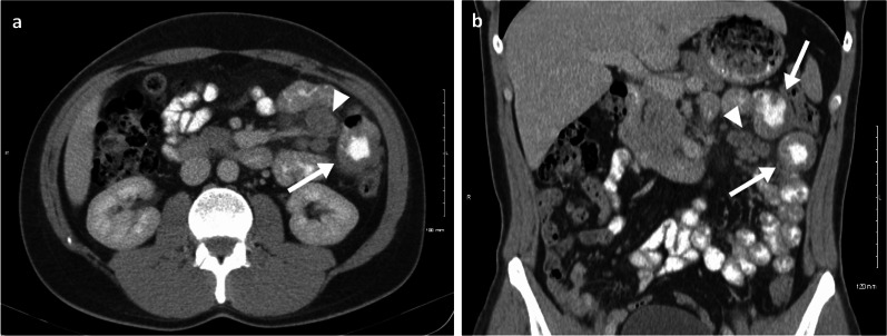
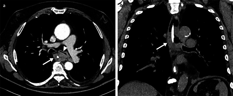
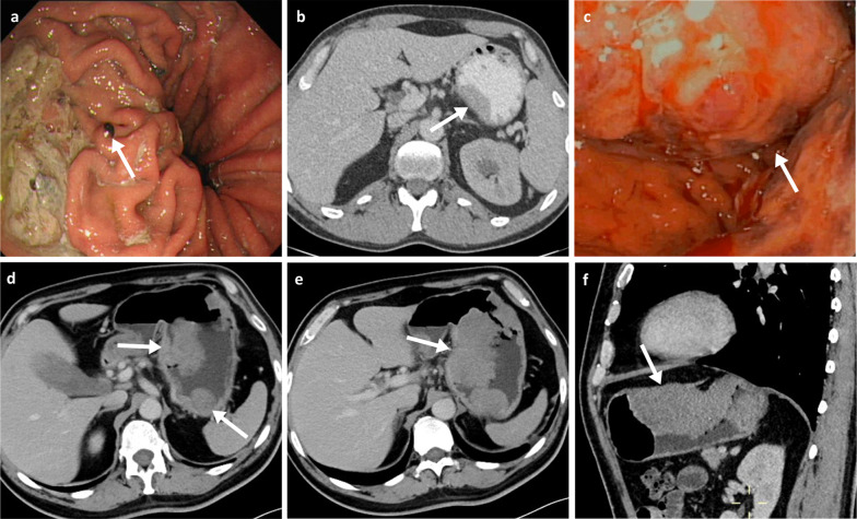
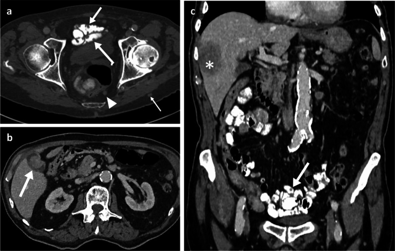
Melanoma is the most aggressive form of skin cancer, with tendency to spread to any organ of the human body, including the gastrointestinal tract (GIT). The diagnosis of metastases to the GIT can be difficult, as they may be clinically silent for somewhile and may occur years after the initial melanoma diagnosis. CT imaging remains the standard modality for staging and surveillance of melanoma patients, and in most cases, it will be the first imaging modality to identify GIT lesions. However, interpretation of CT studies in patients with melanoma can be challenging as lesions may be subtle and random in distribution, as well as sometimes mimicking other conditions. Even so, early diagnosis of GIT metastases is critical to avoid emergency hospitalisations, whilst surgical intervention can be curative in some cases. In this review, we illustrate the various imaging presentations of melanoma metastases within the GIT, discuss the clinical aspects and offer advice on investigation and management. We offer tips intended to aid radiologists in their diagnostic skills and interpretation of melanoma imaging scans.

Keywords: Gastrointestinal tract; Magnetic resonance imaging; Melanoma; Metastases; Tomography (X-ray computed).


Conflict of interest statement

The authors declare no competing interests.

Figures

Fig. 1
Fig. 1
Case report: oral and intravenous portal phase contrast-enhanced CT axial (a) and coronal (b) of a 33-year-old male patient diagnosed with left lower eyelid melanoma which subsequently metastasised to the lymph nodes, lungs, brain, SB and subcutaneous tissue in the following 5-year period. Complete response was achieved after chemotherapy, immunotherapy and radiotherapy. One year after treatment completion he relapsed, presenting with two jejunal metastases (arrows) and involvement of the draining mesenteric lymph nodes (arrow heads). Surgical resection was then performed with no evidence of disease recurrence to date, after 10 years of follow-up
Fig. 2
Fig. 2
Arterial phase contrast-enhanced CT axial (a) and non-enhanced coronal (b) of a 58-year-old male patient demonstrating a large oesophageal metastatic melanoma mass (arrows) with luminal narrowing and compression of the left main bronchus (arrowhead). A nasogastric tube is in situ
Fig. 3
Fig. 3
Gastric melanoma metastases. a Endoscopy of a 69-year-old female patient with a melanotic nodule in the proximal gastric body (arrow). b Oral and intravenous portal phase contrast-enhanced CT axial of a 42-year-old male patient with a sessile soft-tissue melanoma metastasis associated with the lesser curvature (arrow). cf Endoscopy and portal phase contrast-enhanced CT axial and sagittal of a 51-year-old male patient demonstrating a large melanoma metastasis within the body of the stomach with resulting luminal narrowing (arrows)
Fig. 4
Fig. 4
Oral and intravenous portal phase contrast-enhanced CT axial (a, b) and coronal (c) of an 85-year-old male patient with numerous small melanoma metastases throughout the bowel with nodular mucosal thickening (arrows in a, c), metastatic peritoneal nodules (arrowhead in a), gallbladder metastasis (arrow in b), liver metastasis (* in c) and subcutaneous metastasis (thin arrow in a)
Fig. 5
Fig. 5
Oral and intravenous portal phase contrast-enhanced CT axial (a) and coronal (c) of a 70-year-old male patient with an eccentric mural melanoma metastasis within the proximal jejunum (arrows). PET-CT axial (b) and coronal (d) demonstrating FDG avidity with an SUVmax of 21.7 (arrows)
Fig. 6
Fig. 6
Duodenal and small bowel metastases. Portal phase contrast-enhanced CT axial (a) of an 80-year-old male patient with a large aneurysmal metastatic melanoma mass in the duodenum (arrow) and adjacent necrotic node (arrowhead). Oral and intravenous portal phase contrast-enhanced CT axial of a 57-year-old male patient with large jejunal melanoma metastasis (b: arrow) and anterior peritoneal nodule (c: arrow)
Fig. 7
Fig. 7
Portal phase contrast-enhanced CT axial and coronal (a, b) of a 71-year-old male patient demonstrating a large cavitating small bowel melanoma metastasis with urinary bladder tethering and dome invasion (arrows). Further separate caecal pole metastasis (arrowhead)
Fig. 8
Fig. 8
Portal phase contrast-enhanced CT axial (a, b) and oblique coronal (c) of a 71-year-old female patient with ileal melanoma metastasis with resulting obstructing intussusception (arrows)
Fig. 9
Fig. 9
Colonic melanoma metastases. Portal phase contrast-enhanced CT axial (a) of a 71-year-old male patient with a caecal pole melanoma metastasis (arrow). Portal phase contrast-enhanced CT axial (b) and coronal (c) of 71-year-old female patient with a melanoma metastatic deposit to the sigmoid colon with circumferential thickening and enhancement (arrows) and pathological left iliac nodes (arrowhead)
Fig. 10
Fig. 10
Portal phase contrast-enhanced CT sagittal (a) and axial (b), PET-CT sagittal (c) and axial (d), and MRI T2WI sagittal (e) and T2WI axial (f) of a 57-year-old female patient with an anorectal metastatic melanoma (arrows) with a craniocaudal length of 5 cm, situated 1.3 cm from the anal verge with involvement of the right internal sphincter complex
Fig. 11
Fig. 11
Portal phase contrast-enhanced CT axial (a), colonoscopy (b), PET-CT axial (c) and MRI T2WI axial (d) of a 64-year-old male patient with an infiltrative metastatic anorectal melanoma mass. The PET-CT (c) also demonstrates a pathological right inguinal node (arrowhead)
Fig. 12
Fig. 12
Portal phase contrast-enhanced CT axial (a, c) and sagittal (b), MRI T2WI axial (d, f) and T2WI sagittal (e) of a 77-year-old male patient with a metastatic melanoma anal canal mass (arrows) with extensive lymphadenopathy involving the mesorectal fat, iliac, inguinal and retroperitoneal chains (arrowheads)
Fig. 13
Fig. 13
MRI T1WI and T2WI axial (a, b), PET-CT axial (c) and flexible sigmoidoscopy (d) of a 71-year-old female patient with a metastatic melanoma anorectal mass (arrows). Black pigment is visible within the ulcerated part of the lesion on sigmoidoscopy
Fig. 14
Fig. 14
MRI T1WI and T2WI axial (a, b), portal phase contrast-enhanced CT axial (c) and PET-CT axial (obtained two months prior to the shown MRI and CT) (d) of a 57-year-old female patient with a large metastatic melanoma anorectal mass (arrows). The mass demonstrates internal foci of higher T1 signal. PET uptake is also seen within a left inguinal node
Fig. 15
Fig. 15
Peritoneal metastatic melanoma cases. a Oral and intravenous portal phase contrast-enhanced CT axial of 65-year-old left female with paracolic gutter nodules (arrows). b Portal phase contrast-enhanced CT axial of a 45-year-old female patient with a peritoneal deposit adjacent to the hepatic flexure (arrow). c Portal phase contrast-enhanced CT axial of a 73-year-old female patient with a large left upper quadrant necrotic peritoneal mass (arrow). d Portal phase contrast-enhanced CT axial of a 61-year-old female patient with a large left iliac fossa necrotic peritoneal mass (arrow) as well as numerous peritoneal, retroperitoneal and cutaneous nodules/masses
Fig. 16
Fig. 16
Portal phase contrast-enhanced CT axial (a) and coronal (b), and abdominal ultrasound (c) of a 48-year-old male patient with extensive metastatic melanoma peritoneal disease with innumerable nodules and omental cake (arrows)

References

    1. Marks R. Epidemiology of melanoma. Clin Exp Dermatol. 2000;25(6):459–463. doi: 10.1046/j.1365-2230.2000.00693.x. - DOI - PubMed
    1. Henley SJ, HMa J, Jemal A. Annual report to the nation on the status of cancer, part I: National Cancer Statistics. Cancer. 2020;126(10):2225–2249. doi: 10.1002/cncr.32802. - DOI - PMC - PubMed
    1. Saginala K, Barsouk A, Aluru JS, Rawla P, Barsouk A. Epidemiology of melanoma. Med Sci (Basel) 2021;9(4):63. - PMC - PubMed
    1. *SEER*Explorer: An Interactive Website for SEER Cancer Statistics. 7/4/22. https://seer.cancer.gov/explorer.
    1. Ajithkumar T, Parkinson C, Fife K, Corrie P, Jefferies S. Evolving treatment options for melanoma brain metastases. Lancet Oncol. 2015;16(13):e486–e497. doi: 10.1016/S1470-2045(15)00141-2. - DOI - PubMed

LinkOut - more resources