. 2022 Jan-Dec;14(1):2127456.
doi: 10.1080/19490976.2022.2127456.

Bifidobacterium spp. and their metabolite lactate protect against acute pancreatitis via inhibition of pancreatic and systemic inflammatory responses

Affiliations

Bifidobacterium spp. and their metabolite lactate protect against acute pancreatitis via inhibition of pancreatic and systemic inflammatory responses

Han Li et al. Gut Microbes. 2022 Jan-Dec.

Abstract

Severe acute pancreatitis (SAP) is a critical illness characterized by a severe systemic inflammatory response resulting in persistent multiple organ failure and sepsis. The intestinal microbiome is increasingly appreciated to play a crucial role in modulation of AP disease outcome, but limited information is available about the identity and mechanism of action for specific commensal bacteria involved in AP-associated inflammation. Here we show that Bifidobacteria, particularly B. animalis, can protect against AP by regulating pancreatic and systemic inflammation in germ-free (GF) and oral antibiotic-treated (Abx) mouse models. Colonization by B. animalis and administration of its metabolite lactate protected Abx and GF mice from AP by reducing serum amylase concentration, ameliorating pancreatic lesions and improving survival rate after retrograde injection of sodium taurocholate. B. animalis relieved macrophage-associated local and systemic inflammation of AP in a TLR4/MyD88- and NLRP3/Caspase1-dependent manner through its metabolite lactate. Supporting our findings from the mouse study, clinical AP patients exhibited a decreased fecal abundance of Bifidobacteria that was inversely correlated with the severity of systemic inflammatory responses. These results may shed light on the heterogeneity of clinical outcomes and drive the development of more efficacious therapeutic interventions for AP, and potentially for other inflammatory disorders.

Keywords: Bifidobacterium; immunomodulation; lactate; macrophages; microbial metabolite; pancreatic and systemic inflammation.


Conflict of interest statement

No potential conflict of interest was reported by the author(s).

Figures
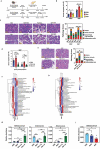
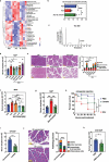
Figure 1.
Figure 1.
Intensive short-term antibiotic treatment via oral gavage exacerbates caerulein-induced acute pancreatitis. (a) Schematic illustration of experiments in MAP and SAP models induced by caerulein injection, including PBS, antibiotic (Abx) treatment and fecal microbiota transplantation (FMT). (b) Serum amylase levels and (c) representative H&E staining of pancreatic sections with pathology scores for SAP or MAP mice at 12 h post-treatment (hpt) with PBS, Abx or FMT (n = 5 each). (d) Serum amylase activity of MAP mice treated with PBS, Abx oral gavage (o.g.) or Abx in drinking water (i.w.) at 12 or 24 hpt (n = 10 each). (e) Pathological sections and pancreatic pathology scores from mice from (d) at 24 hpt. (f) The microbiota composition in fecal samples collected from groups treated with PBS, Abx o.g. or Abx i.w. was analyzed at the genus level by 16S rDNA sequencing (n = 3–5). (g) The relative abundance of Bifidobacterium, Akkermansia and Enterocococcus in fecal samples collected from (f). (h) 16S rDNA sequencing analysis from fecal samples of PBS control, MAP and SAP mice (n = 3–5). (i) Relative abundance of Bifidobacterium from (h). Data are from two independent experiments, and P values were determined by unpaired two-tailed Student’s t-test; *: P < .05; **: P < .01; ***: P < .001; ****: P < .0001.
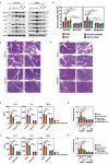
Figure 2.
Figure 2.
Colonization by Bifidobacterium spp. is protective against MAP and SAP. (a) Compositional analysis of Bifidobacterium genus in fecal samples collected from MAP mice administered Abx via driving water (n = 4). Figure depicts the top 20 Bifidobacterium spp. detected by deep sequencing. The percentage indicates the proportion of the bacteria in the total intestinal flora. (b) Serum amylase concentration of MAP mice treated with PBS, Abx, or fecal microbiota transplantation (FMT), or Abx-treated mice colonized with B. pseudocatenulatum, B. animalis, B. adolescentis or Enterococcus faecalis at 12 h post MAP modeling. (c) Pancreatic histopathology and pathology scores from mice treated with PBS, Abx or Abx-treated mice colonized with B. animalis or E. faecalis at 12 hpt of MAP (n = 5). (d) Serum amylase activity from SAP mice treated with PBS, Abx or Abx-treated mice colonized by B. animalis at 12 hpt (n = 5). (e) Serum amylase concentration in SAP mice treated with PBS, Abx or Abx-treated mice colonized with different doses of B. animalis. (f) Survival kinetics in surgical SAP model. Survival was observed for 72 h following retrograde injection of 5% sodium taurocholate into the pancreatic-bile duct in mice treated with PBS, Abx or Abx-treated mice colonized by B. animalis or E. faecalis (n = 6–14). (g) Serum amylase level of mice from (f) at 6 h post-retrograde injection. (h) Serum amylase activity in uncolonized or B. animalis-colonized GF mice at 12 h following SAP modeling (n = 5). (i) Pancreatic histopathological changes and pathology scores of mice from (h). (j) Serum amylase activity of conventionally raised SPF mice (CNV), either treated with PBS or colonized by B. animalis at 12 h post-SAP induction (n = 5). Data are from two independent experiments, and P values were determined by unpaired two-tailed Student’s t-test. Broken lines indicate the limit of detection (LD) of the assay. *: P < .05; **: P < .01; ***: P < .001; ****: P < .0001; ns: not significant.
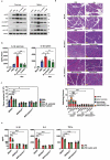
Figure 3.
Figure 3.
B. animalis colonization and exogenous lactate administration protect the host from MAP and SAP. (a) Heatmap showing differential metabolites in serum of individual animals treated with PBS, Abx or Abx-treated mice colonized by B. animalis at 12 h post-SAP induction. (b) Absolute lactate concentration in serum samples collected from mice from (a). (c) The supernatant of B. animalis cultures was analyzed by liquid chromatography coupled with tandem mass spectrometry (LC-MS/MS). (d) Serum amylase concentration, (e) pancreatic histopathological changes and pathology scores of mice treated with PBS, Abx or Abx-treated mice administered either lactate (0.24 g/kg), acetate, propionate or butyrate at 12 h post-induction of MAP (n = 6–10). (f) Serum amylase activity in mice treated with PBS, Abx or Abx-treated mice given different doses of lactate at 12 h post-induction of MAP. (g) Serum amylase level of mice treated with PBS, Abx or Abx-treated mice receiving lactate gavage (0.24 g/kg) at 12 h post-induction of SAP (n = 5). (h) Survival kinetics in surgical SAP model. Survival was observed for 72 h following retrograde injection of 5% sodium taurocholate into the pancreatic-bile duct in mice treated with PBS, Abx or Abx-treated mice receiving lactate gavage (0.24 g/kg) (n = 13–14). (i) Serum amylase level, (j) histopathological changes and pancreatic pathology scores in untreated- or lactate-treated germ-free (GF) mice at 12 h post-induction of SAP (n = 5). (k) Serum amylase activity of conventionally raised SPF mice (CNV) treated with PBS or lactate (0.24 g/kg) at 12 h post-induction of SAP (n = 5). Data are from two independent experiments, and P values were determined by unpaired two-tailed Student’s t-test. Broken lines indicate the limit of detection (LD). *: P < .05; **: P < .01; ***: P < .001; ****: P < .0001; ns: not significant.
Figure 4.
Figure 4.
B. animalis and its metabolite lactate protect against AP by suppressing macrophage-mediated pancreatic and systemic inflammatory responses. (a) Frequency of pancreatic macrophages and neutrophils in mice treated with PBS, Abx, or Abx-treated mice either colonized by B. animalis or given lactate gavage (0.24 g/kg) at 12 h post-induction of SAP (n = 5). (b) Representative immunofluorescence images of necrotic areas within the pancreas of SAP mice treated with PBS, Abx or Abx-treated mice receiving fecal microbiota transplantation (FMT), colonization by B. animalis or E. faecalis, or lactate gavage (0.24 g/kg). In addition, germ-free (GF) mice were colonized by B. animalis or treated with lactate (0.24 g/kg) at 12 h post-induction of SAP (n = 5). Macrophages were double-stained with CD11b (green) and F4/80 (red) antibodies. (c, g) Frequency of CD68-expressing M1 macrophages (left panel) or M1/M2 macrophage ratio (right panel) in the (c) pancreas or (g) spleen of mock or SAP mice treated with PBS, Abx, or Abx-treated mice colonized by B. animalis or given lactate gavage at 12 h post-induction (n = 5). (d, h) Relative mRNA expression of Il1b, Il6, Il10 and Tnfa in the (d) pancreas or (h) spleen of mock or SAP mice treated with PBS, Abx, or Abx-treated mice colonized by B. animalis or given lactate gavage at 12 h post-induction (n = 5–6). Fold change is relative to respective mock mice. (e) Frequency of monocytes in PBMCs and (f) serum expression (ELISA) of IL-1β, IL-6 and TNF-α in mock or SAP mice treated with PBS, Abx or Abx-treated mice colonized by B. animalis or given lactate gavage (0.24 g/kg) at 12 h post-induction (n = 5). Data are from two independent experiments, and P values were determined by unpaired two-tailed Student’s t-test. *: P < .05; **: P < .01; ***: P < .001; ****: P < .0001.
Figure 5.
Figure 5.
Relief of macrophage-mediated inflammation during AP by B. animalis colonization or lactate administration requires TLR4-MyD88 signaling. (a) Representative western blot showing expression of NF-κB signaling pathway-related molecules in the pancreas and spleen of mice treated with PBS, Abx, or Abx-treated mice colonized by B. animalis or given lactate gavage at 12 h post-induction of SAP (All experiments were repeated independently three times. one representative experiment was shown.) (b) Serum amylase activity, (c, d) pancreatic tissue lesions and (g, h) pathology scores of Tlr4−/− or Myd88−/− mice or their respective wild-type (WT) parent strain, treated with PBS, Abx or Abx-treated mice colonized by B. animalis or given lactate gavage at 12 h post-induction of SAP (n = 5). Expression (ELISA) of IL-1β, IL-6 and TNF-α in peritoneal macrophages extracted from (e) Tlr4−/− or (f) Myd88−/− mice or their respective WT parents. Cells were pretreated or not with lactic acid (15 mM), and subsequently stimulated with LPS (200 ng/mL). Data are from two independent experiments, and P values were determined by unpaired two-tailed Student’s t-test. *: P < .05; **: P < .01; ***: P < .001; ****: P < .0001.
Figure 6.
Figure 6.
Suppression of NLRP3 inflammasome-mediated inflammatory responses is required for B. animalis- and lactate-dependent protection against AP. (a) Western blot showing expression of molecules related to the NLRP3 signaling pathway and (b) ELISA showing secretion of IL-1β in the pancreas and spleen of mice treated with PBS, Abx or Abx-treated mice colonized by B. animalis or given lactate gavage at 12 h post-induction of SAP. (c) Serum amylase concentration, (d) histopathological changes and pathology scores in the pancreas of wild-type (WT), Nlrp3−/− or Caspase1−/− mice treated with PBS, Abx or Abx-treated mice colonized by B. animalis or given lactate gavage (0.24 g/kg) at 12 h post-induction of SAP (n = 5). (e) Expression (ELISA) of IL-1β, IL-6 and TNF-α in peritoneal macrophages extracted from WT, Nlrp3−/− or Caspase1−/− mice. Cells were pretreated or not with lactic acid (15 mM), and subsequently stimulated with LPS (200 ng/mL). Data are from two independent experiments, and P values were determined by unpaired two-tailed Student’s t-test. *: P < .05; **: P < .01; ***: P < .001; ****: P < .0001.
Figure 7.
Figure 7.
The decreased fecal abundance of Bifidobacterium in patients with acute pancreatitis inversely correlates with the severity of their systemic inflammatory responses. (a) The microbiota composition of human fecal samples collected from healthy volunteers (HC) and patients with clinical MAP, MSAP or SAP was analyzed at the genus level by 16S rDNA sequencing. (b) Comparison of relative abundance of Bifidobacterium between HC and AP patients (left panel) or among groups of patients with different levels of AP severity (right panel). (c) Serum lactate concentration in patients with AP. Differential serum expression of IL-1β (d) and IL-6 (e) between HC and AP patients (left panel) or among groups of patients with different levels of AP severity (right panel), as determined by ELISA. Correlation between fecal Bifidobacterium relative abundance and serum concentration of IL-1β (f) or IL-6 (g) in AP patients. Statistical significance was evaluated by one-way ANOVA. Data are from two independent experiments, and P values were determined by unpaired two-tailed Student’s t-test. *: P < .05; **: P < .01; ***: P < .001; ****: P < .0001; ns: not significant.

References

    1. Sendler M, Maertin S, John D, Persike M, Weiss FU, Krüger B, Wartmann T, Wagh P, Halangk W, Schaschke N, et al. Cathepsin B activity initiates apoptosis via digestive protease activation in pancreatic acinar cells and experimental pancreatitis. J Biol Chem. 2016;291(28):14717–23. doi:10.1074/jbc.M116.718999. - DOI - PMC - PubMed
    1. Lerch MM, Saluja AK, Dawra R, Saluja M, Steer ML.. The effect of chloroquine administration on two experimental models of acute pancreatitis. Gastroenterology. 1993;104(6):1768–1779. doi:10.1016/0016-5085(93)90658-Y. - DOI - PubMed
    1. Criddle DN, McLaughlin E, Murphy JA, Petersen OH, Sutton R. The pancreas misled: signals to pancreatitis. Pancreatology. 2007;7(5–6):436–446. doi:10.1159/000108960. - DOI - PubMed
    1. Jakkampudi A, Jangala R, Reddy BR, Mitnala S, Reddy DN, Talukdar R. NF-kappaB in acute pancreatitis: mechanisms and therapeutic potential. Pancreatology. 2016;16(4):477–488. doi:10.1016/j.pan.2016.05.001. - DOI - PubMed
    1. Gukovskaya AS, Gukovsky I, Algul H, Habtezion A. Autophagy, inflammation, and immune dysfunction in the pathogenesis of pancreatitis. Gastroenterology. 2017;153(5):1212–1226. doi:10.1053/j.gastro.2017.08.071. - DOI - PMC - PubMed

Publication types

MeSH terms