Review
. 2022 Sep 29:10:e14053.
doi: 10.7717/peerj.14053. eCollection 2022.

Macrophages play a key role in tissue repair and regeneration

Affiliations
Review

Macrophages play a key role in tissue repair and regeneration

Yajie Yu et al. PeerJ. .

Abstract

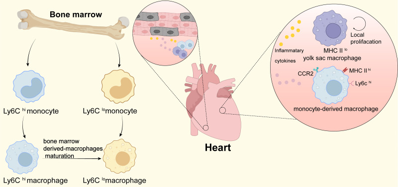
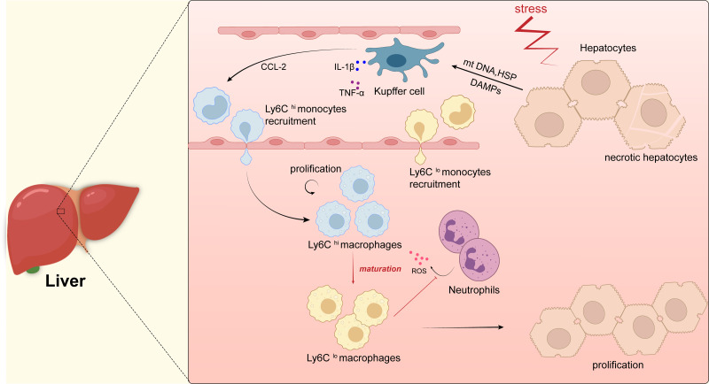
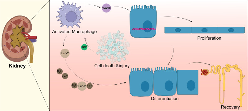
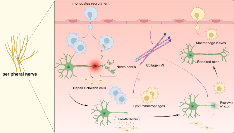
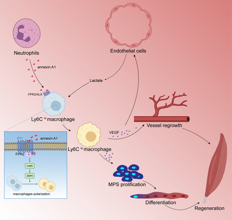
Tissue regeneration after body injury has always been a complex problem to resolve for mammals. In adult mammals, the repair process after tissue injury is often accompanied by continuous and extensive fibrosis, which leads to scars. This process has been shown to severely hinder regeneration. Macrophages, as widely distributed innate immune cells, not only play an important role in various pathological processes, but also participate in the repair process before tissue regeneration and coordinate the regeneration process after repair. This review will discuss the various forms and indispensability of macrophages involved in repair and regeneration, and how macrophages play a role in the repair and regeneration of different tissues.

Keywords: Cytokine; Polarization; Regeneration; Tissue repair; Macrophage.


Conflict of interest statement

The authors declare there are no competing interests.

Figures

Figure 1
Figure 1. Macrophages in heart.
This figure shows the origin of infiltrating monocytes in the bone marrow after tissue injury. Ly6Chi macrophages can mature and differentiate into Ly6Clo macrophages after infiltrating tissues. In the heart at homeostasis, tissue-resident macrophages are the predominant macrophage type, which originate from the yolk sac and are low in expression of MHC II (MHC IIlo); during periods of stress in the heart, the number of cardiac macrophages increases significantly through local proliferation and monocyte recruitment. CCR2+ monocyte-derived macrophages and heart-resident macrophages produce and secrete large amounts of pro-inflammatory cytokines that promote cardiomyocyte regeneration and contribute to cardiac tissue remodeling.
Figure 2
Figure 2. Macrophages in liver.
Kupffer cells are immobilized within the lumen of liver sinusoidal endothelial cells (LSECs) and when hepatocytes are damaged, intracellular components, such as mitochondrial DNA (mtDNA) or heat shock proteins (HSP), are released as dangerously relevant molecular patterns (DAMP). DAMPs activate Kupffer cells, which in turn secrete inflammatory cytokines (e.g., TNF- α, IL-1 β, etc.) as well as CCL-2. CCL-2 recruits Ly6Chi monocytes from the blood to infiltrate tissues and differentiate into pro-inflammatory macrophages (Ly6Chi), which will mature into Ly6Clo macrophages with restorative and anti-inflammatory properties when inflammation resolves, they promote the regeneration of liver parenchymal cells and thus the repair of the liver. In addition, Ly6Clo macrophages inhibit the secretion of reactive oxygen species (ROS) by neutrophils.
Figure 3
Figure 3. Macrophages in kidney.
Acute kidney injury can lead to tubular epithelial cell death and injury, resulting in macrophage recruitment and activation. Activated macrophages promote the activation of the Wnt pathway, as shown by the upregulation of Wnt7b in macrophages, which directs the proliferation of renal tubular epithelial cells; macrophages also secrete lipid transport protein 2(LCN-2), and apoptotic cells secrete sphingosine-1-phosphate to promote the secretion of more Lcn-2 by macrophages, which binds to iron ions for transport to epithelial cells and promotes epithelial cell differentiation.
Figure 4
Figure 4. Macrophages in peripheral nerve.
After peripheral nerve injury, monocytes are recruited to the site of injury and differentiate into pro-inflammatory macrophages, macrophages phagocytose myelin debris, and Schwann cells isolate and dedifferentiate into repairing Schwann cells, which secrete cytokines to promote axonal regeneration while the pro-inflammatory macrophages mature into repairing macrophages (Ly6Clo). The polarization of macrophages stimulates the secretion of collagen VI from the membranes of the peripheral nerve, which induces macrophage polarization by positive feedback and participates in axonal repair. The repairing Schwann cells guide axonal regeneration and the repairing macrophages release anti-inflammatory cytokines. After complete nerve regeneration, the macrophages withdraw from the peripheral nerve tissue.
Figure 5
Figure 5. Macrophages are involved in skeletal muscle regeneration.
Skeletal muscle is a highly vascularized tissue and endothelial cells are the main cell type of blood vessels. During muscle regeneration, endothelial cells release lactic acid, which directly controls macrophage function. Under lactic acid stimulation, pro-inflammatory macrophages differentiate into reparative macrophages, which secrete large amounts of vascular endothelial growth factor (VEGF), actively supporting muscle vascularization and forming a positive feedback pathway. VEGF also induces the proliferation and differentiation of myogenic progenitor cells (MPCs) in skeletal muscle. Muscle regeneration depends on the self-renewal of MPCs to replenish the muscle stem cell pool, and MPCs repair damaged muscle by differentiating and fusing with each other; following injury to skeletal muscle, membrane linked protein A1 (Annexin A1), which is secreted in large numbers by neutrophils, activates the FPR2/ALX receptor on the surface of macrophages and the downstream adenylate-activated protein kinase signaling (AMPK) pathway, promoting the conversion of pro-inflammatory macrophages to reparative macrophages.

References

    1. Agah A, Kyriakides TR, Lawler J, Bornstein P. The lack of thrombospondin-1 (TSP1) dictates the course of wound healing in double-TSP1/TSP2-null mice. American Journal of Pathology. 2002;161:831–839. doi: 10.1016/s0002-9440(10)64243-5. - DOI - PMC - PubMed
    1. Allen RE, Boxhorn LK. Inhibition of skeletal muscle satellite cell differentiation by transforming growth factor-beta. Journal of Cellular Physiology. 1987;133:567–572. doi: 10.1002/jcp.1041330319. - DOI - PubMed
    1. Antoniades CG, Quaglia A, Taams LS, Mitry RR, Hussain M, Abeles R, Possamai LA, Bruce M, McPhail M, Starling C, Wagner B, Barnardo A, Pomplun S, Auzinger G, Bernal W, Heaton N, Vergani D, Thursz MR, Wendon J. Source and characterization of hepatic macrophages in acetaminophen-induced acute liver failure in humans. Hepatology. 2012;56:735–746. doi: 10.1002/hep.25657. - DOI - PubMed
    1. Aurora AB, Porrello ER, Tan W, Mahmoud AI, Hill JA, Bassel-Duby R, Sadek HA, Olson EN. Macrophages are required for neonatal heart regeneration. Journal of Clinical Investigation. 2014;124:1382–1392. doi: 10.1172/JCI72181. - DOI - PMC - PubMed
    1. Baeck C, Wehr A, Karlmark KR, Heymann F, Vucur M, Gassler N, Huss S, Klussmann S, Eulberg D, Luedde T, Trautwein C, Tacke F. Pharmacological inhibition of the chemokine CCL2 (MCP-1) diminishes liver macrophage infiltration and steatohepatitis in chronic hepatic injury. Gut. 2012;61:416–426. doi: 10.1136/gutjnl-2011-300304. - DOI - PubMed

Publication types

LinkOut - more resources