. 2023 Jan 9;13(1):56-69.
doi: 10.1158/2159-8290.CD-22-0381.

A Small Molecule Reacts with the p53 Somatic Mutant Y220C to Rescue Wild-type Thermal Stability

Affiliations

A Small Molecule Reacts with the p53 Somatic Mutant Y220C to Rescue Wild-type Thermal Stability

Keelan Z Guiley et al. Cancer Discov. .

Abstract

The transcription factor and tumor suppressor protein p53 is the most frequently mutated and inactivated gene in cancer. Mutations in p53 result in deregulated cell proliferation and genomic instability, both hallmarks of cancer. There are currently no therapies available that directly target mutant p53 to rescue wild-type function. In this study, we identify covalent compsounds that selectively react with the p53 somatic mutant cysteine Y220C and restore wild-type thermal stability.

Significance: The tumor suppressor p53 is the most mutated gene in cancer, and yet no therapeutics to date directly target the mutated protein to rescue wild-type function. In this study, we identify the first allele-specific compound that selectively reacts with the cysteine p53 Y220C to rescue wild-type thermal stability and gene activation. See related commentary by Lane and Verma, p. 14. This article is highlighted in the In This Issue feature, p. 1.


Figures

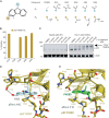
Figure 1. Development of a covalent PhiKan083. A, Chemical structures of the carbazole series. B, 1 μmol/L p53 Y220C-CL was incubated with 10 μmol/L of the carbazole compounds at 4°C for 24 hours. Adduct formation was observed by LC/MS. C, HepG2 (p53WT/+) or Huh-7 (p53 Y220C/−) cells were treated with the indicated KG5 concentration for 1 hour at 37°C and target engagement was observed by gel shift following a copper click reaction with TAMRA-azide. D, Crystal structure of p53 Y220C-CL bound to KG3 at 2.4 Å resolution. E, Crystal structure of the p53 Y220C-PhiKan083 complex PDB 2VUK (8).
Figure 1.
Development of a covalent PhiKan083. A, Chemical structures of the carbazole series. B, 1 μmol/L p53 Y220C-CL was incubated with 10 μmol/L of the carbazole compounds at 4°C for 24 hours. Adduct formation was observed by LC/MS. C, HepG2 (p53WT/+) or Huh-7 (p53 Y220C/−) cells were treated with the indicated KG5 concentration for 1 hour at 37°C and target engagement was observed by gel shift following a copper click reaction with TAMRA-azide. D, Crystal structure of p53 Y220C-CL bound to KG3 at 2.4 Å resolution. E, Crystal structure of the p53 Y220C–PhiKan083 complex (Protein Data Bank 2VUK; ref. 8).
Figure 2. A methacrylic indole compound induced a novel p53 conformational state. A, Chemical structures of methacrylic indole compound series. B, 1 μmol/L p53 Y220C-CL was incubated with 10 μmol/L indole series at 4°C for 24 hours. Adduct formation was analyzed by LC/MS. C, Crystal structure of p53 Y220C-CL bound to KG6 in a novel conformational state at 1.6 Å resolution. D, Crystal structure of p53 Y220C-CL bound to KG10 at 2.0 Å resolution.
Figure 2.
A methacrylic indole compound induced a novel p53 conformational state. A, Chemical structures of methacrylic indole compound series. B, 1 μmol/L p53 Y220C-CL was incubated with 10 μmol/L indole series at 4°C for 24 hours. Adduct formation was analyzed by LC/MS. C, Crystal structure of p53 Y220C-CL bound to KG6 in a novel conformational state at 1.6 Å resolution. D, Crystal structure of p53 Y220C-CL bound to KG10 at 2.0 Å resolution.
Figure 3. Azaindole compounds stabilized p53 Y220C to WT levels. A, Chemical structures of the azaindole series. B, p53 Y220C-CL was fully labeled by the compounds, purified by size-exclusion chromatography, and its thermal stability was analyzed by differential scanning fluorimetry. The difference in Tm relative to apo p53 Y220C-CL is displayed. C, Crystal structure of p53 Y220C-CL bound to KG13 at 1.7 Å resolution. D, 1 μmol/L of p53 Y220C-CL or p53 Y220C-CL D228A were incubated with 10 μmol/L compound at 4°C for 24 hours. Adduct formation was analyzed by LC/MS.
Figure 3.
Azaindole compounds stabilized p53 Y220C to WT levels. A, Chemical structures of the azaindole series. B, p53 Y220C-CL was fully labeled by the compounds and purified by size-exclusion chromatography, and its thermal stability was analyzed by differential scanning fluorimetry. The difference in Tm relative to unliganded p53 Y220C-CL is displayed. C, Crystal structure of p53 Y220C-CL bound to KG13 at 1.7 Å resolution. D, 1 μmol/L of p53 Y220C-CL or p53 Y220C-CL D228A was incubated with 10 μmol/L compound at 4°C for 24 hours. Adduct formation was analyzed by LC/MS.
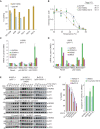
Figure 4. KG13 stabilized cellular p53 Y220C and activated target gene expression. A, 1 μmol/L of p53 WT or p53 Y220C was incubated with 10 μmol/L of compound at 4°C for 24 hours. Adduct formation was analyzed by LC/MS. B, Cellular thermal shift assay intensity values were plotted with calculated Tagg values where the KG13-treated sample showed an increase in thermal stability of cellular p53 Y220C compared with DMSO control. C, RT-qPCR results for 25 μmol/L KG13-treated H1299 cells stably expressing p53 R273C or Y220C. D, RT-qPCR results for 10 μmol/L Nutlin-3a or 25 μmol/L KG13-treated U2OS cells stably expressing p53 Y220C. E, Western blot for DMSO, 10 μmol/L Nutlin-3a, and 10 μmol/L KG13-treated cell panel. F, Viability and caspase-3/7 activity for NUGC-4, NUGC-3, or BxPC-3 cells treated with KG13.
Figure 4.
KG13 stabilized cellular p53 Y220C and activated target gene expression. A, 1 μmol/L of p53 WT or p53 Y220C was incubated with 10 μmol/L of compound at 4°C for 24 hours. Adduct formation was analyzed by LC/MS. B, CETSA intensity values were plotted with calculated Tagg values, where the KG13-treated sample showed an increase in thermal stability of cellular p53 Y220C compared with DMSO control. C, RT-qPCR results for 25 μmol/L KG13-treated H1299 cells stably expressing p53 R273C or Y220C. rel., relative. D, RT-qPCR results for 10 μmol/L Nutlin-3a (Nutlin) or 25 μmol/L KG13-treated U2OS cells stably expressing p53 Y220C. E, Western blot for DMSO, 10 μmol/L Nutlin-3a, and 10 μmol/L KG13-treated cell panel. F, Viability and caspase-3/7 activity for NUGC-4, NUGC-3, or BxPC-3 cells treated with KG13.

Comment in

  • Covalent Rescue of Mutant p53.
    Lane DP, Verma CS. Lane DP, et al. Cancer Discov. 2023 Jan 9;13(1):14-16. doi: 10.1158/2159-8290.CD-22-1212. Cancer Discov. 2023. PMID: 36620883

References

    1. Alexandrova EM, Mirza SA, Xu S, Schulz-Heddergott R, Marchenko ND, Moll UM. P53 loss-of-heterozygosity is a necessary prerequisite for mutant p53 stabilization and gain-of-function in vivo. Cell Death Dis 2017;8:e2661–5. - PMC - PubMed
    1. McBride KA, Ballinger ML, Killick E, Kirk J, Tattersall MHN, Eeles RA, et al. . Li-Fraumeni syndrome: cancer risk assessment and clinical management. Nat Rev Clin Oncol 2014;11:260–71. - PubMed
    1. Boettcher S, Miller PG, Sharma R, McConkey M, Leventhal M, Krivtsov AV, et al. . A dominant-negative effect drives selection of TP53 missense mutations in myeloid malignancies. Science 2019;365:599–604. - PMC - PubMed
    1. Ventura A, Kirsch DG, McLaughlin ME, Tuveson DA, Grimm J, Lintault L, et al. . Restoration of p53 function leads to tumour regression in vivo. Nature 2007;445:661–5. - PubMed
    1. Xue W, Zender L, Miething C, Dickins RA, Hernando E, Krizhanovsky V, et al. . Senescence and tumour clearance is triggered by p53 restoration in murine liver carcinomas. Nature 2007;445:656–60. - PMC - PubMed

Publication types