Review
. 2022 Dec;20(6):516-531.
doi: 10.1007/s11914-022-00757-4. Epub 2022 Oct 5.

CSF1R as a Therapeutic Target in Bone Diseases: Obvious but Not so Simple

Affiliations
Review

CSF1R as a Therapeutic Target in Bone Diseases: Obvious but Not so Simple

David A Hume et al. Curr Osteoporos Rep. 2022 Dec.

Abstract

Purpose of review: The purpose of the review is to summarize the expression and function of CSF1R and its ligands in bone homeostasis and constraints on therapeutic targeting of this axis.

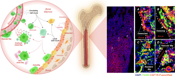
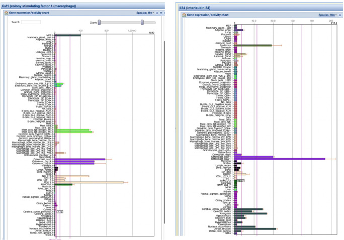
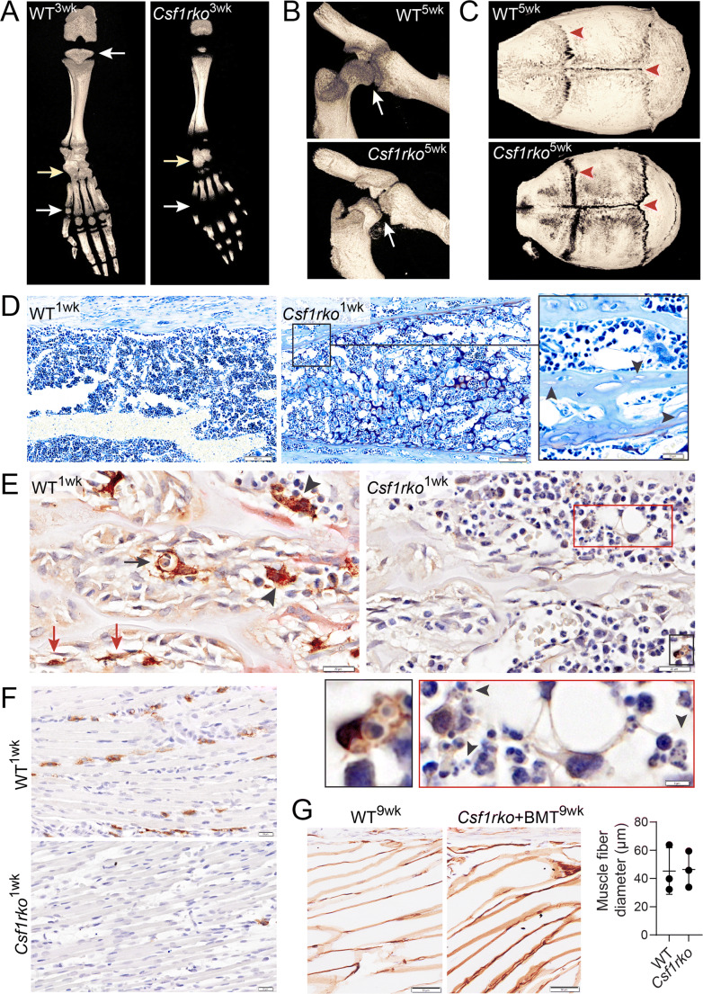
Recent findings: Bone development and homeostasis depends upon interactions between mesenchymal cells and cells of the mononuclear phagocyte lineage (MPS), macrophages, and osteoclasts (OCL). The homeostatic interaction is mediated in part by the systemic and local production of growth factors, macrophage colony-stimulating factor (CSF1), and interleukin 34 (IL34) that interact with a receptor (CSF1R) expressed exclusively by MPS cells and their progenitors. Loss-of-function mutations in CSF1 or CSF1R lead to loss of OCL and macrophages and dysregulation of postnatal bone development. MPS cells continuously degrade CSF1R ligands via receptor-mediated endocytosis. As a consequence, any local or systemic increase or decrease in macrophage or OCL abundance is rapidly reversible. In principle, both CSF1R agonists and antagonists have potential in bone regenerative medicine but their evaluation in disease models and therapeutic application needs to carefully consider the intrinsic feedback control of MPS biology.

Keywords: CSF1R; Homeostasis; Macrophage; Osteoclast; Osteoporosis.


Conflict of interest statement

The authors have no existing conflicts of interest.

Figures

Fig. 1
Fig. 1
The mononuclear phagocyte populations of mouse bone. The schematic at left highlights the diversity of mononuclear phagocyte populations (osteoclasts, monocyte, macrophages (Mϕ), and committed progenitors (CMP, OMP)) found in mouse bone and the local and systemic sources of growth factors CSF1 and IL34. The images at right show the co-localization of the Csf1r-FusionRed transgene [•] and the macrophage-restricted F4/80 antigen [•]. The low power image confirms that the majority of bone marrow hematopoietic and mesenchymal (osteoblast, fibroblast, adipocyte, endothelial) cells lack expression of the FusionRed reporter [•]. Osteoclasts (Panel A) express FusionRed but lack F4/80 [•]. Mϕ populations associated with the bone surface (B), hematopoietic islands (C), and sinusoids (D) express both FusionRed and F4/80. F4/80 is relatively low on monocytes. Panels A and D contain multiple FusionRed-positive mononuclear cells; presumptive monocytes; and their progenitors. At bottom of Panel D, the large FusionRed-positive, F4/80-negative cell is a megakaryocyte
Fig. 2
Fig. 2
Expression of Csf1 and Il34 mRNA in mouse. Figure shows screenshots from BioGPS.org of the expression of Csf1 and Il34 mRNA in a wide range of mouse tissues and cells. Csf1 is expressed in embryonic fibroblasts (MEF), ES cells, stimulated mast cells, osteoblasts, and various mesenchymal cell lines. As expected, Il34 was detected primarily in the epidermis and brain, but was also induced during differentiation of primary osteoblasts
Fig. 3
Fig. 3
Phenotypic analysis of skeletal development in Csf1r knockout rats. Homozygous knockout mutation of Csf1r in rats (Csf1rko) was generated by Pridans et al. [•] and impacts on skeletal development were described in [•]. This figure contains previously unpublished images highlighting aspects of the mutant phenotype and its reversal by bone marrow cell transfer. AC show μCT images demonstrating the delay in ossification in the digits and formation of the hip-joint and defective cranial suture closure. D shows the tibial diaphyseal region in juvenile WT and Csf1rko rats and the presence of empty osteocyte lacunae. The Csf1rko was associated with substantive loss of both OCL pink stained for expression of tartrate-resistant acid phosphatase (TRAP) and osteomacs (brown stained for expression of IBA1 (E). The inset shows that the residual macrophages showed evidence of efferocytosis but extracellular pyknotic nuclei are also evident (arrowheads). F shows that IBA1+ macrophages are also depleted in skeletal muscle in the Csf1rko rat associated with the reduction in muscle fiber diameter and postnatal somatic growth retardation that is observed in these animals [79]. Transfer of WT bone marrow (BMT) at weaning without conditioning corrected all of these musculoskeletal phenotypes, as exemplified by restoration of muscle mass and fiber diameter (G)

References

    1. Wiktor-Jedrzejczak W, Bartocci A, Ferrante AW, Jr, Ahmed-Ansari A, Sell KW, Pollard JW, Stanley ER. Total absence of colony-stimulating factor 1 in the macrophage-deficient osteopetrotic (op/op) mouse. Proc Natl Acad Sci U S A. 1990;87(12):4828–4832. doi: 10.1073/pnas.87.12.4828. - DOI - PMC - PubMed
    1. Yoshida H, Hayashi S, Kunisada T, Ogawa M, Nishikawa S, Okamura H, et al. The murine mutation osteopetrosis is in the coding region of the macrophage colony stimulating factor gene. Nature. 1990;345(6274):442–444. doi: 10.1038/345442a0. - DOI - PubMed
    1. Van Wesenbeeck L, Odgren PR, MacKay CA, D’Angelo M, Safadi FF, Popoff SN, et al. The osteopetrotic mutation toothless (tl) is a loss-of-function frameshift mutation in the rat Csf1 gene: evidence of a crucial role for CSF-1 in osteoclastogenesis and endochondral ossification. Proc Natl Acad Sci U S A. 2002;99(22):14303–14308. doi: 10.1073/pnas.202332999. - DOI - PMC - PubMed
    1. Lajeunesse D, Busque L, Menard P, Brunette MG, Bonny Y. Demonstration of an osteoblast defect in two cases of human malignant osteopetrosis. Correction of the phenotype after bone marrow transplant. J Clin Invest. 1996;98(8):1835–1842. doi: 10.1172/JCI118984. - DOI - PMC - PubMed
    1. Sobacchi C, Schulz A, Coxon FP, Villa A, Helfrich MH. Osteopetrosis: genetics, treatment and new insights into osteoclast function. Nat Rev Endocrinol. 2013;9(9):522–536. doi: 10.1038/nrendo.2013.137. - DOI - PubMed

Publication types

MeSH terms

Substances