. 2022 Oct 5;18(10):e1009967.
doi: 10.1371/journal.pgen.1009967. eCollection 2022 Oct.

The RNA helicase DDX6 controls early mouse embryogenesis by repressing aberrant inhibition of BMP signaling through miRNA-mediated gene silencing

Affiliations

The RNA helicase DDX6 controls early mouse embryogenesis by repressing aberrant inhibition of BMP signaling through miRNA-mediated gene silencing

Jessica Kim et al. PLoS Genet. .

Abstract

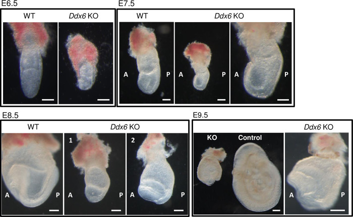
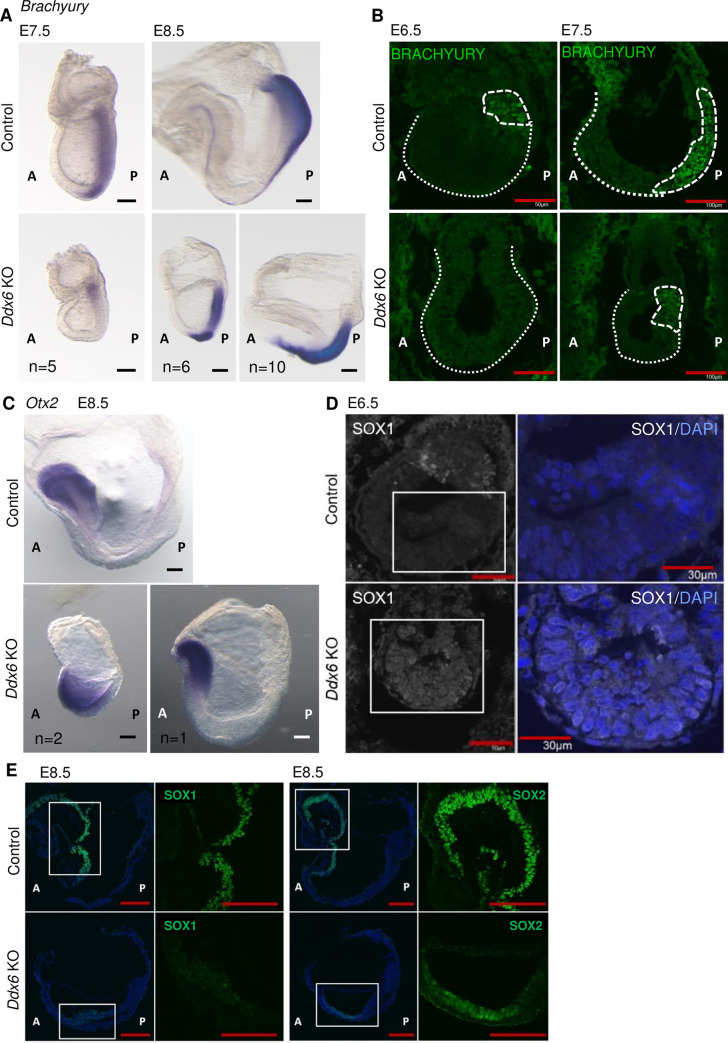
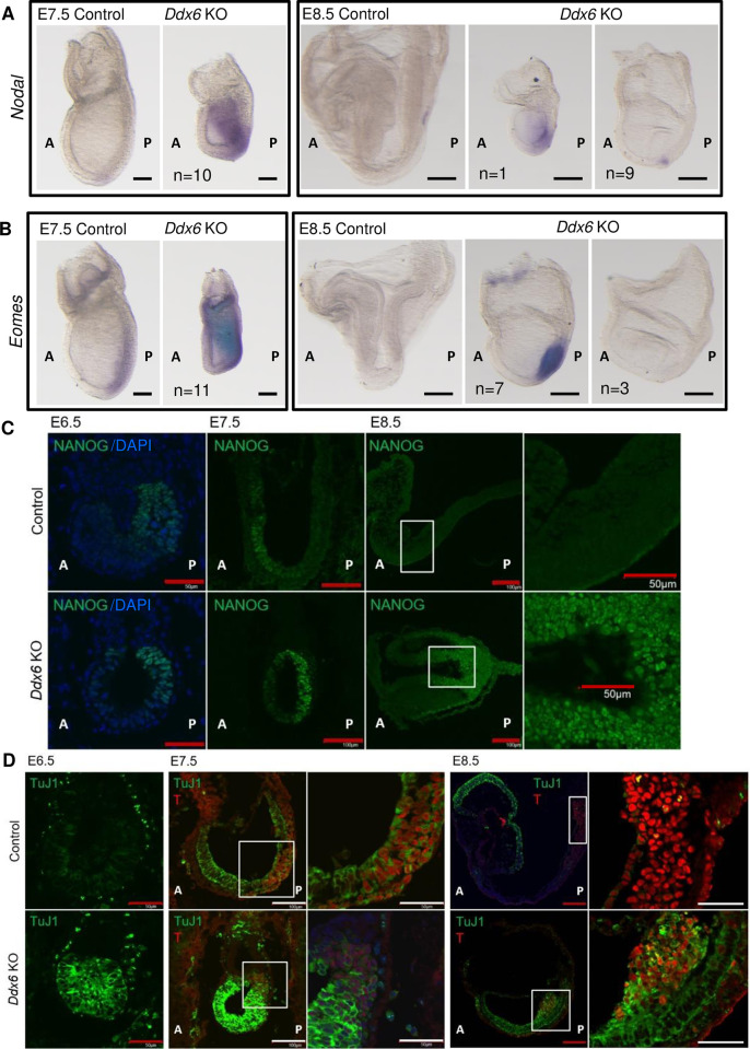
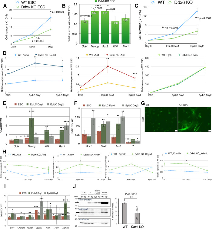
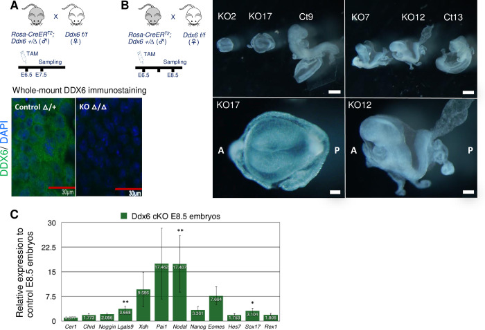
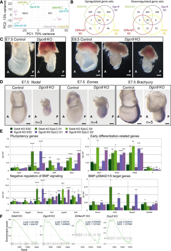
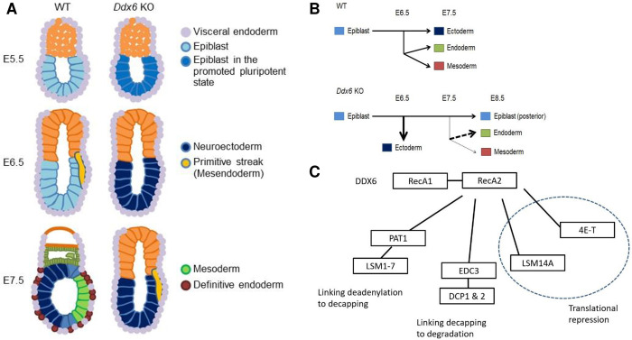
The evolutionarily conserved RNA helicase DDX6 is a central player in post-transcriptional regulation, but its role during embryogenesis remains elusive. We here show that DDX6 enables proper cell lineage specification from pluripotent cells by analyzing Ddx6 knockout (KO) mouse embryos and employing an in vitro epiblast-like cell (EpiLC) induction system. Our study unveils that DDX6 is an important BMP signaling regulator. Deletion of Ddx6 causes the aberrant upregulation of the negative regulators of BMP signaling, which is accompanied by enhanced expression of Nodal and related genes. Ddx6 KO pluripotent cells acquire higher pluripotency with a strong inclination toward neural lineage commitment. During gastrulation, abnormally expanded Nodal and Eomes expression in the primitive streak likely promotes endoderm cell fate specification while inhibiting mesoderm differentiation. We also genetically dissected major DDX6 pathways by generating Dgcr8, Dcp2, and Eif4enif1 KO models in addition to Ddx6 KO. We found that the miRNA pathway mutant Dgcr8 KO phenocopies Ddx6 KO, indicating that DDX6 mostly works along with the miRNA pathway during early development, whereas its P-body-related functions are dispensable. Therefore, we conclude that DDX6 prevents aberrant upregulation of BMP signaling inhibitors by participating in miRNA-mediated gene silencing processes. Overall, this study delineates how DDX6 affects the development of the three primary germ layers during early mouse embryogenesis and the underlying mechanism of DDX6 function.


Conflict of interest statement

The authors have declared that no competing interests exist.

Figures

Fig 1
Fig 1. Ddx6/ embryos exhibited growth delay and morphological defects.
Pictures of embryos prepared from E6.5-E9.5 littermates. At E8.5, two morphologically distinct mutant embryos are shown; earlier than head-fold formation (1) and with head-fold structure (2). The right panels of E7.5 and E9.5 are the magnified images of Ddx6/ embryos in the left panels. (Scale: 100 μm for E6.5 and E7.5; 200 μm for E8.5 and E9.5).
Fig 2
Fig 2. Ddx6/ embryos display developmental defects: mesoderm formation failure and premature neural induction.
(A) Whole-mount ISH of E7.5 & E8.5 embryos with a Brachyury probe (Scale: 100 μm, n = 5 for E7.5; 200 μm, n = 16 for E8.5). (B) E6.5 & E7.5 embryo frozen section IHC for BRACHYURY (Scale: 50 μm for E6.5, n = 2; 100 μm for E7.5, n = 3). Embryo parts are indicated by dotted lines. (C) Whole-mount ISH of E8.5 embryos with an Otx2 probe (Scale: 100 μm, n = 3). (D) E6.5 embryo frozen section IHC for SOX1 (Scale: 50 μm for lower magnification, 30 μm for higher magnification, n = 2). Square parts are enlarged in the right panels. Shown with DAPI staining. (E) E8.5 embryo frozen section IHC for SOX1 & SOX2 (Scale: 100 μm, n = 3).
Fig 3
Fig 3. Ddx6/ embryos exhibit defects that are associated with increased Nodal expression.
(A-B) Whole-mount ISH of E7.5 & E8.5 embryos with (A) a Nodal probe (Scale: 100 μm, n = 10 for E7.5; 200 μm, n = 10 for E8.5) and (B) an Eomes probe (Scale: 100 μm, n = 11 for E7.5; 200 μm, n = 10 for E8.5). Expressions of Nodal and Eomes were maintained in E8.5 Ddx6 KO embryos, which were younger than the head-fold stage (left), but down-regulated as in WT at the head-fold stage (right). (C) E6.5~E8.5 embryo frozen section IHC for NANOG (Scale: 50 μm for E6.5, n = 2; 100 μm for E7.5, n = 3; 100 μm for lower magnification of E8.5, 50 μm for higher magnification, n = 3). Square parts are enlarged in the right panels. (D) TuJ1 expression in the epiblast. E6.5~E8.5 embryo frozen section IHC for TuJ1 & T (BRACHYURY). The signal intensity is comparable only between the same embryonic day samples. (Scale: 50 μm for E6.5, n = 2; 100 μm for lower magnification, 50 μm for higher magnification, n = 3 for E7.5 & E8.5). Square parts are enlarged in the right panels.
Fig 4
Fig 4. BMP signaling is repressed in Ddx6/ pluripotent cells.
(A) Cell counting of ESCs over a three-day culture period. Mean ± SEM. Significance was calculated by the Student’s t-test (n = 5). (B) qRT-PCR examining the relative expression of pluripotency markers in Ddx6/ ESCs to WT ESCs. Mean ± SEM. Student’s t-test (n = 7~9). (C) Cell counting during the ESC-to-EpiLC induction period. Mean ± SEM. Student’s t-test (n = 13 for WT, n = 7 for Ddx6△/△). (D) qRT-qPCR examining the expression pattern of Nodal, Fgf5, and Zic3. Mean ± SEM. Student’s t-test (n = 9 for Nodal & Fgf5, n = 7 for Zic3) (*p ≤ 0.05, **p ≤ 0.01, ***p ≤ 0.001, ****p ≤ 0.0001). (E-F, I) qRT-PCR analysis of the expression trend of several key genes during the EpiLC induction period. Each bar represents the relative expression of Ddx6/ cells to WT cells at the indicated time point. Mean ± SEM. Student’s t-test. (E) Major pluripotency genes (n = 7~9). (F) Early neuroectoderm and mesendoderm lineage markers (n = 7~9). (I) The negative regulators of BMP signaling (n = 6~9). (G) TuJ1 ICC (immunocytochemistry) on Day1 of monolayer differentiation (Scale: 50 μm) (n = 3). (H) qRT-PCR analysis of the expression of the known BMP-SMAD1/5 target genes in ESCs. Each value is the relative expression to WT ESCs (n = 5). (J) Western blot for endogenous SMAD5 and phosphorylated SMAD1/5 in mESCs. Four conditions: LDN (LDN193189: BMP Type I receptor ALK2/3 inhibitor)/—(non-treated)/ BMP4 1/10 (1/10 dilution of rBMP4 treated sample)/ BMP4 (rBMP4 treated). The quantified signal intensity of the band is displayed on the right-side graph (n = 6).
Fig 5
Fig 5. Conditional knockout of Ddx6 quickly upregulates expression of the BMP signaling inhibitors and Nodal.
(A) Tamoxifen was injected at E6.5. Whole-mount DDX6 immunostaining confirmed that the complete depletion of DDX6 takes approximately 1 day (Scale: 30 μm, n = 3). DDX6 in green, DAPI in blue. (B) E8.5 cKO embryos exhibited similar phenotypes to conventional KO embryos (Scale: 500 μm for group, 100 μm for KO17, 200 μm for KO12) (n = 6). (C) qRT-PCR analysis of several key genes in Ddx6 cKO E8.5 embryos. Embryos exhibiting similar morphology to the KO12 were used for analysis. Mean ± SEM. Significance was calculated by the Wilcoxon rank-sum test (n = 9~10) (* α = 0.05 significance level, ** α = 0.01).
Fig 6
Fig 6. Genetic dissection of the DDX6 functions indicates that the DDX6-miRNA pathway is essential during early embryogenesis.
(A) PCA plot of the ESC and EpiLC Day2 samples of each genotype group. (B) Summary of the GSEA results of ESC RNA-seq data. The gene sets that were changed in Ddx6 KO, Dgcr8 KO, Eif4enif1 KO, and Dcp2 KO were compared. (C) Dgcr8 KO embryos exhibited similar morphological defects to Ddx6 KO embryos at E7.5, but they became more malformed at E8.5 (Scale: 100 μm for E7.5; 200 μm for E8.5). (D) Whole-mount ISH of E7.5 Dgcr8 KO embryos with Nodal, Eomes, and Brachyury probes (Scale: 100 μm, n = 3 for Nodal, n = 4 for Eomes, n = 5 for Brachyury). (E) Comparison of gene expression between Ddx6 KO and Dgcr8 KO. qRT-PCR analysis of several key genes during the EpiLC induction period. Each bar represents the relative expression of KO cells to WT cells at the indicated time point. Mean ± SEM. Student’s t-test (n ≥ 3) (*p ≤ 0.05, **p ≤ 0.01, ***p ≤ 0.001, ****p ≤ 0.0001). (F) GSEA enrichment plot of the “negative regulation of BMP signaling pathway” gene set in four mutant ESCs. Black bars represent the position of the genes that belong to this gene set (n = 45) in the whole ranked gene list. The green line shows the overall distribution of this gene set (whether over-represented at the top (left) or bottom (right) of the ranked list of genes).
Fig 7
Fig 7. Schemes showing developmental defects caused by loss of DDX6-mediated RNA regulation.
(A) Development of the three primary embryonic germ layers is largely affected by DDX6. Neuroectoderm is specified earlier than WT, whereas formation of the primitive streak is delayed (The smaller size of the Ddx6 mutant is not reflected in the images). (B) Changes in cell-lineage specification from pluripotent stem cells caused by Ddx6 loss are depicted on a horizontal diagram. Uncommitted Ddx6/ pluripotent cells possess promoted pluripotency and strongly favor commitment to the neuronal lineage. In WT embryos, the mesendoderm lineage arises at ~E6.5 as the primitive streak is formed, and three germ layers simultaneously develop at ~E7.5. In Ddx6 KO embryos, premature neural induction occurs with a one-day delay in primitive streak formation. During mesendoderm segregation, definitive endoderm specification is increased, whereas mesoderm specification is greatly reduced due to the patterning defect of the primitive streak. Posterior epiblast cells cannot exit pluripotency on time, impeding the differentiation processes. (C) The interaction of DDX6 with other gene silencing effector protein complexes.

Similar articles

Cited by

References

    1. Kaikkonen MU, Lam MTY, Glass CK. Non-coding RNAs as regulators of gene expression and epigenetics. Vol. 90, Cardiovascular Research. 2011. doi: 10.1093/cvr/cvr097 - DOI - PMC - PubMed
    1. Dassi E. Handshakes and fights: The regulatory interplay of RNA-binding proteins. Vol. 4, Frontiers in Molecular Biosciences. 2017. doi: 10.3389/fmolb.2017.00067 - DOI - PMC - PubMed
    1. Tanner NK, Linder P. DExD/H box RNA helicases: From generic motors to specific dissociation functions. Vol. 8, Molecular Cell. 2001. doi: 10.1016/s1097-2765(01)00329-x - DOI - PubMed
    1. Jankowsky E. RNA helicases at work: Binding and rearranging. Vol. 36, Trends in Biochemical Sciences. 2011. doi: 10.1016/j.tibs.2010.07.008 - DOI - PMC - PubMed
    1. Linder P, Jankowsky E. From unwinding to clamping-the DEAD box RNA helicase family. Vol. 12, Nature Reviews Molecular Cell Biology. 2011. doi: 10.1038/nrm3154 - DOI - PubMed

Publication types