. 2023:16:22-37.
doi: 10.1109/RBME.2022.3212038. Epub 2023 Jan 5.

Recent Advances in Biosensors for Detection of COVID-19 and Other Viruses

Recent Advances in Biosensors for Detection of COVID-19 and Other Viruses

Shobhit K Patel et al. IEEE Rev Biomed Eng. 2023.

Abstract

This century has introduced very deadly, dangerous, and infectious diseases to humankind such as the influenza virus, Ebola virus, Zika virus, and the most infectious SARS-CoV-2 commonly known as COVID-19 and have caused epidemics and pandemics across the globe. For some of these diseases, proper medications, and vaccinations are missing and the early detection of these viruses will be critical to saving the patients. And even the vaccines are available for COVID-19, the new variants of COVID-19 such as Delta, and Omicron are spreading at large. The available virus detection techniques take a long time, are costly, and complex and some of them generates false negative or false positive that might cost patients their lives. The biosensor technique is one of the best qualified to address this difficult challenge. In this systematic review, we have summarized recent advancements in biosensor-based detection of these pandemic viruses including COVID-19. Biosensors are emerging as efficient and economical analytical diagnostic instruments for early-stage illness detection. They are highly suitable for applications related to healthcare, wearable electronics, safety, environment, military, and agriculture. We strongly believe that these insights will aid in the study and development of a new generation of adaptable virus biosensors for fellow researchers.


Figures
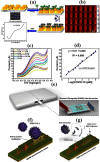
Fig. 1.
Fig. 1.
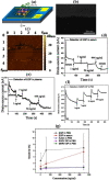
Various COVID-19 detecting biosensors (a) Illustrative prototype of COVID-19 electrochemical micro-biosensor including immobilization of antibody and obtaining COVID-19 protein (b) AFM topography of the gold assembly (c) SWV voltammograms for different compositions of COVID-19 protein from 5000 fmol L-1 0.1 formula imagemol L-1 after interacting antiCOVID-19 electrode (d) the relation among the composition of COVID-19 protein and oxidation current peak (Reprinted from , copyright Springer Nature) (e) A POC biosensor based on biomedical waveguide (BiMW) interferometric technology (f) Detection of intact SARS-CoV-2 using BiMW with CONVAT technique (g) Viral genomic analysis using BiMW with CONVAT technique (Reprinted from , copyright IOPSCIENCE).
Fig. 2.
Fig. 2.
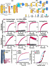
Electrochemical biosensor-based nucleic acid testing (a) Brief workflow of the biosensor with N genes and S genes RCA, SEM images (b) Silica core, silica-methylene blue (SiMB), and silica-acridine orange (SiAO) with various diameter size represented as mean value with standard deviation (c) Comparison of step-wise and one-step sandwich hybridization process with N gene as the target, No significant difference is observed in these two strategies with sensitivity test for N and S genes positive correlation for a current response, increment in differential pulse voltammetry as N and S gene concentration increases (d) Numerous sequence position of N genes and S genes target orders with mismatch and non-complementary target orders. Dark areas and underlined bases show the non-complementary arrangements to the target gene and mismatch bases, respectively (e) The identification of N genes and S genes in 55 cDNA specimens equated with the Cq result from qRT-PCR (N genes (Blue dots), S genes (Orange dots)) (Reprinted from , copyright Springer Nature).
Fig. 3.
Fig. 3.
(a) Illustrative mechanism of the proposed sensor (b) The thermodynamics behind the activation of sensor (c) Specificity of the proposed sensor for various cognate targets and targets for other biosensors (d) Incorporating of de novo SARS-CoV-2 RBD binder, Increment in luminescence due to the addition of trimeric spike protein, and detection over a range of analyte compositions in buffer (Reprinted from , copyright Springer Nature).
Fig. 4.
Fig. 4.
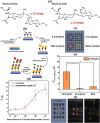
Label-free, Multiplex microarray biosensor for influenza detection (a) Human and avian cell surface sialyloligosaccharide structures (b) Preparation and incubation of AIR microarray chips for virus-glycan binding detection (c) Glycan-based receptor analogue microarray layout design (d) AIR glycosyl microarray responses to H1N1pdm and H13N8 viruses at various viral doses (e) The glycan microarray's quantitative response data to the H1N1pdm virus (left) and the H13N8 virus at right. (f) Analysis protocol for AIR images (Reprinted from , copyright ACS Publications).
Fig. 5.
Fig. 5.
(a) Illustrative diagram of reduced graphene oxide-based FET biosensor (b) SEM image of reduced Graphene Oxide sheet (c) AFM used to determine the depth of the reduced Graphene Oxide sheet, which was found to be less than 2 nm (d) EGP inserted in formula image × human serum (e) EGP inserted in formula image × human plasma (f) Detection of MARV GP in PBS (g) Sensitivity as a function of protein composition is investigated. (Reprinted from , copyright Springer Nature).
Fig. 6.
Fig. 6.
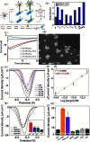
Gold nanoparticles based electrochemical biosensor based Zika virus detection (a) Illustration of the concept to amplify the nanobioconjugate-based signal (b) Assessment of molar ration using anti-Dig-HRP as a reporter (c) For various gold and NaCl composition, chronoamperometry curves for the gold electrodeposition (d) SEM images of gold resultant structures at various enlargement (e) DPV plots of nanobioconjugates accumulated onto SPGEs and (f) Resultant calibration curve (g) DPV plots of three positive ra serum specimens (h) Matrix effect and specificity (Reprinted from , copyright Springer Nature).

Similar articles

Cited by

References

    1. Ye Z. W., Yuan S., Yuen K. S., Fung S. Y., Chan C. P., and Jin D. Y., “Zoonotic origins of human coronaviruses,” Int. J. Biol. Sci., vol. 16, no. 10. pp. 1686–1697, 2020, doi: 10.7150/ijbs.45472. - DOI - PMC - PubMed
    1. Weaver S. C. et al., “Zika virus: History, emergence, biology, and prospects for control,” Antiviral Res., vol. 130. pp. 69–80, 2016, doi: 10.1016/j.antiviral.2016.03.010. - DOI - PMC - PubMed
    1. Weyer J., Grobbelaar A., and Blumberg L., “Ebola virus disease: History, epidemiology and outbreaks,” Curr. Infect. Dis. Rep., vol. 17, no. 5. pp. 1–8, 2015, doi: 10.1007/s11908-015-0480-y. - DOI - PubMed
    1. Dick G. W. A., “Zika Virus (I), Isolations and serological specificity,” Trans. R. Soc. Trop. Med. Hyg., vol. 46, no. 5, pp. 509–520, 1952, doi: 10.1016/0035-9203(52)90042-4. - DOI - PubMed
    1. MacNamara F. N., “Zika virus: A report on three cases of human infection during an epidemic of jaundice in Nigeria,” Trans. R. Soc. Trop. Med. Hyg., vol. 48, no. 2, pp. 139–145, 1954, doi: 10.1016/0035-9203(54)90006-1. - DOI - PubMed

Publication types