. 2022 Oct 4;41(1):111448.
doi: 10.1016/j.celrep.2022.111448.

Molecular basis for processing of topoisomerase 1-triggered DNA damage by Apn2/APE2

Affiliations

Molecular basis for processing of topoisomerase 1-triggered DNA damage by Apn2/APE2

Jessica S Williams et al. Cell Rep. .

Abstract

Topoisomerase 1 (Top1) incises DNA containing ribonucleotides to generate complex DNA lesions that are resolved by APE2 (Apn2 in yeast). How Apn2 engages and processes this DNA damage is unclear. Here, we report X-ray crystal structures and biochemical analysis of Apn2-DNA complexes to demonstrate how Apn2 frays and cleaves 3' DNA termini via a wedging mechanism that facilitates 1-6 nucleotide endonucleolytic cleavages. APN2 deletion and DNA-wedge mutant Saccharomyces cerevisiae strains display mutator phenotypes, cell growth defects, and sensitivity to genotoxic stress in a ribonucleotide excision repair (RER)-defective background harboring a high density of Top1-incised ribonucleotides. Our data implicate a wedge-and-cut mechanism underpinning the broad-specificity Apn2 nuclease activity that mitigates mutagenic and genome instability phenotypes caused by Top1 incision at genomic ribonucleotides incorporated by DNA polymerase epsilon.

Keywords: APE2; Apn2; CP: molecular biology; DNA damage; DNA repair; DNA replication; S. cerevisiae; Top1; X-ray crystallography; ribonucleotide.


Conflict of interest statement

Declaration of interests The authors declare no competing interests.

Figures
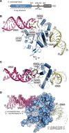
Figure 1.
Figure 1.. X-ray crystal structure of the Apn2-DNA complex
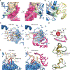
(A) Domain architecture of Apn2. (B) Crystal structure of the N-terminal EEP domain of S. cerevisiae Apn2 bound to DNA with 1 of the 4 Apn2 protomers in the asymmetric unit of the DNA complex crystal represented. (C) An orthogonal view of the catalytic domain structure bound to DNA. (D) A surface representation of the Apn2-DNA complex with the electron density map of the DNA overlaid (final 2Fo-Fc, 1.0 σ at 2.73 Å).
Figure 2.
Figure 2.. Apn2 DNA interactions
(A) Surface representations of the DNA, the WL, and Y33 reveal tight shape complementarity. The 180° rotation shows how Y33, together with the WL, interrogates the minor groove of incoming DNA, making van der Waals contacts with the DNA backbone and a charge interaction with the side chain of N317, stabilizing both the DNA and the WL. (B) Basic surface comprised residues K201, R205, R208, and R273 (green surfaces and sticks) create a groove that recognizes the backbone of the second DNA molecule in the crystal. (C) The WL changes conformation upon DNA binding. The conformational change of the WL allows for new side chain contacts to recognize the DNA substrate, essentially splaying open the strands to make available the damaged 3′ end for cleavage. The WL appears to “bloom,” with conserved residues W312 and R319 serving as the central anchor point out from which the other residues unfurl. The WL tilts down with the DNA interaction to allow N317 to begin to intercalate between the strands and to position K316 for direct interaction with the 3′ DNA termini to be processed. (D) Simplified illustrated model of a fully “bloomed” WL. (E) Orthogonal views of (C). (F) Overlay of ribbon representations of DNA-free and DNA-bound WLs in all Apn2 protomers from both the DNA-free and DNA-present crystal structures.
Figure 3.
Figure 3.. Apn2 nuclease processes a variety of 3′ DNA termini
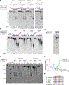
(A) Denaturing gel electrophoresis of dsDNA oligonucleotide substrates carrying a 3′ hydroxyl, 3′ G:A mismatch, 3′ ribouridine (rU), or terminal 2′,3′-cyclic phosphate (see Table S2 for substrates) incubated with recombinant S. cerevisiae maltose binding protein (MBP)-Apn2, PCNA, or MBP as controls. Yellow, position of the FAM label; numbers, oligonucleotide lengths. Gels shown are representative of 3 technical replicates. (B) Denaturing gel electrophoresis exonuclease assays of WT, Y33E, R205E, and R208E mutant Apn2 in the presence and absence of PCNA with the 3′ hydroxyl substrate (see Table S2 for substrates). Gels shown are representative of 3 technical replicates. (C) Denaturing gel electrophoresis of salt-sensitivity exonuclease time course assays of WT and WL deletion mutants in the absence of PCNA. Gels shown are representative of 3 technical replicates. (D) Denaturing gel electrophoresis of salt-sensitivity exonuclease time course assays of WT, WL deletion, and catalytically dead Apn2 in the presence of PCNAwith a 30 hydroxyl substrate (3′OH) (Table S2). Gels shown are representative of 3 technical replicates. (E) Quantification of the band intensities observed in the 10 mM NaCl, 60 min lanes in (C) using Fiji. Experiments are representative example of gels run 3 times. (F) Sequence alignment of the WL with anchor residues highlighted in red and pink circles denoting DNA interaction. The deletion mutation used is indicated.
Figure 4.
Figure 4.. The WL guides endonucleolytic cleavage events
(A) A 3’ FAM-labeled substrate with increasing, non-hydrolyzable phosphorothioate bonds at the 3’ end demonstrate a difference in PCNA-stimulated endonucleolytic cleavage pattern between WT Apn2 and the WL deletion mutant. DNA ladders were produced by T7 (lane 3) or lambda exonuclease (lane 4) digestion of substrate, and by direct synthesis 5-nt 3′-FAM-labeled DNA ladder (lanes 5, 20, 21, and 36). Gels shown are representative of 3 technical replicates. (B) Model of Apn2 recognizing and processing DNA substrate.
Figure 5.
Figure 5.. Apn2 is important for suppressing mutagenesis and promoting normal cell growth and genotoxin resistance in the presence of a high density of Top1-incised nascent leading strand ribonucleotides
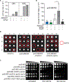
(A) Δ2 to 5-bp mutation rates for the URA3-OR1 (orientation 1) and URA3-OR2 reporter genes in the WT, rnh201Δ, and rnh201Δ apn2Δ mutants were calculated as the proportion of each type of event among the total mutants sequenced, multiplied by the overall mutation rate (Table S4). (B) Tetrad analysis of an APN2/apn2::natMX diploid in a homozygous POL2 or pol2-M644G strain ± RNH201 and TOP1. Plates were photographed after 3 days of growth at 30°C. (C) Deleting APN2 in the pol2-M644G RNase H2-defective strains impairs growth and causes genotoxin sensitivity that is reduced upon the deletion of TOP1. Serial (10-fold) dilutions of cells were plated on rich medium ± HU and photographed after 3 days’ growth at 30°C. (D) Overall spontaneous mutation rates for yeast strains expressing pol2-M644G ± RNH201, APN2, and TOP1 were determined by fluctuation analysis using a URA3 reporter assay. The rate of mutation conferring resistance to 5-FOA was determined as ura3 mutants are resistant to 5-FOA. The median rate ± the 95% confidence interval is displayed. *p = 0.0028. N represents the number of mutation rate replicates and is as follows: pol2-M644G: 57, pol2-M644G apn2Δ: 36, pol2-M644G rnh201Δ: 24, pol2-M644G rnh201Δ apn2Δ: 46, and pol2-M644G rnh201Δ apn2Δ top1Δ: 32.
Figure 6.
Figure 6.. Apn2 prevents base-base mismatch mutagenesis caused by Top1 cleavage at ribonucleotides
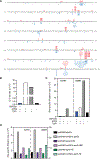
(A) The coding strand of the 804-bp URA3 gene is shown (OR1). Sequence changes observed in independent ura3 mutants are depicted in red above the coding sequence for the pol2-M644G rnh201Δ strain (n = 126; Nick McElhinny et al., 2010) and in blue beneath the coding sequence for the pol2-M644G rnh201Δ apn2Δ strain (n = 191). Letters indicate single-base substitutions, closed triangles indicate single-base additions, open triangles indicate single-base deletions, and short lines indicate 2–5 bp deletions. (B) Δ2- to 5-bp mutation rates in the pol2-M644G ± RNH201, APN2, and TOP1 were calculated as the proportion of each type of event among the total mutants sequenced, multiplied by the overall mutation rate (Table S6). (C) As in (B), but for the A286T and A686T transversion hotspots in the pol2-M644G strain ± RNH201, APN2, and TOP1. (D) As in (C), but for the Apn2 DNA-binding mutants.
Figure 7.
Figure 7.. Overall model of Apn2 processing of 3’ DNA ends produced when Top1 cleaves at genomic ribonucleotides
A model depicting the consequences of Top1 cleavage at unrepaired ribonucleotides incorporated by Pol ε during DNA replication. Processing of a Top1-induced nick can be repaired via mutagenic or error-free repair pathways. Results in yeast demonstrate the importance of Apn2 for promoting cell growth and HU resistance and preventing single-base mismatch mutagenesis in a strain with a high density of unrepaired ribonucleotides (pol2-M644G RNase H2-defective). As these phenotypes are dependent on the presence of Top1, this suggests that the “dirty” 3’ DNA end produced by Top1 incision at a ribonucleotide requires Apn2 for processing and resolution. This may involve multiple enzymatic functions possessed by Apn2, including 3’ exonucleolytic, unwinding, and endonuclease activities.

References

    1. Adams PD, Afonine PV, Bunkó czi G, Chen VB, Davis IW, Echols N, Headd JJ, Hung LW, Kapral GJ, Grosse-Kunstleve RW, et al. (2010). PHENIX: a comprehensive Python-based system for macromolecular structure solution. Acta Crystallogr. D Biol. Crystallogr 66, 213–221. 10.1107/S0907444909052925. - DOI - PMC - PubMed
    1. Allen-Soltero S, Martinez SL, Putnam CD, and Kolodner RD (2014). A saccharomyces cerevisiae RNase H2 interaction network functions to suppress genome instability. Mol. Cell Biol 34, 1521–1534. 10.1128/MCB.00960-13. - DOI - PMC - PubMed
    1. Álvarez-Quilón A, Wojtaszek JL, Mathieu MC, Patel T, Appel CD, Hustedt N, Rossi SE, Wallace BD, Setiaputra D, Adam S, et al. (2020). Endogenous DNA 3’ blocks are vulnerabilities for BRCA1 and BRCA2 deficiency and are reversed by the APE2 nuclease. Mol. Cell 78, 1152–1165.e8. 10.1016/j.molcel.2020.05.021. - DOI - PMC - PubMed
    1. Andres SN, Schellenberg MJ, Wallace BD, Tumbale P, and Williams RS (2015). Recognition and repair of chemically heterogeneous structures at DNA ends. Environ. Mol. Mutagen 56, 1–21. 10.1002/em.21892. - DOI - PMC - PubMed
    1. Burgers PMJ, and Kunkel TA (2017). Eukaryotic DNA replication fork. Annu. Rev. Biochem 86, 417–438. 10.1146/annurev-biochem-061516-044709. - DOI - PMC - PubMed

Publication types

MeSH terms