. 2022 Oct 4;34(10):1578-1593.e6.
doi: 10.1016/j.cmet.2022.09.004.

Single-cell dissection of the obesity-exercise axis in adipose-muscle tissues implies a critical role for mesenchymal stem cells

Affiliations

Single-cell dissection of the obesity-exercise axis in adipose-muscle tissues implies a critical role for mesenchymal stem cells

Jiekun Yang et al. Cell Metab. .

Abstract

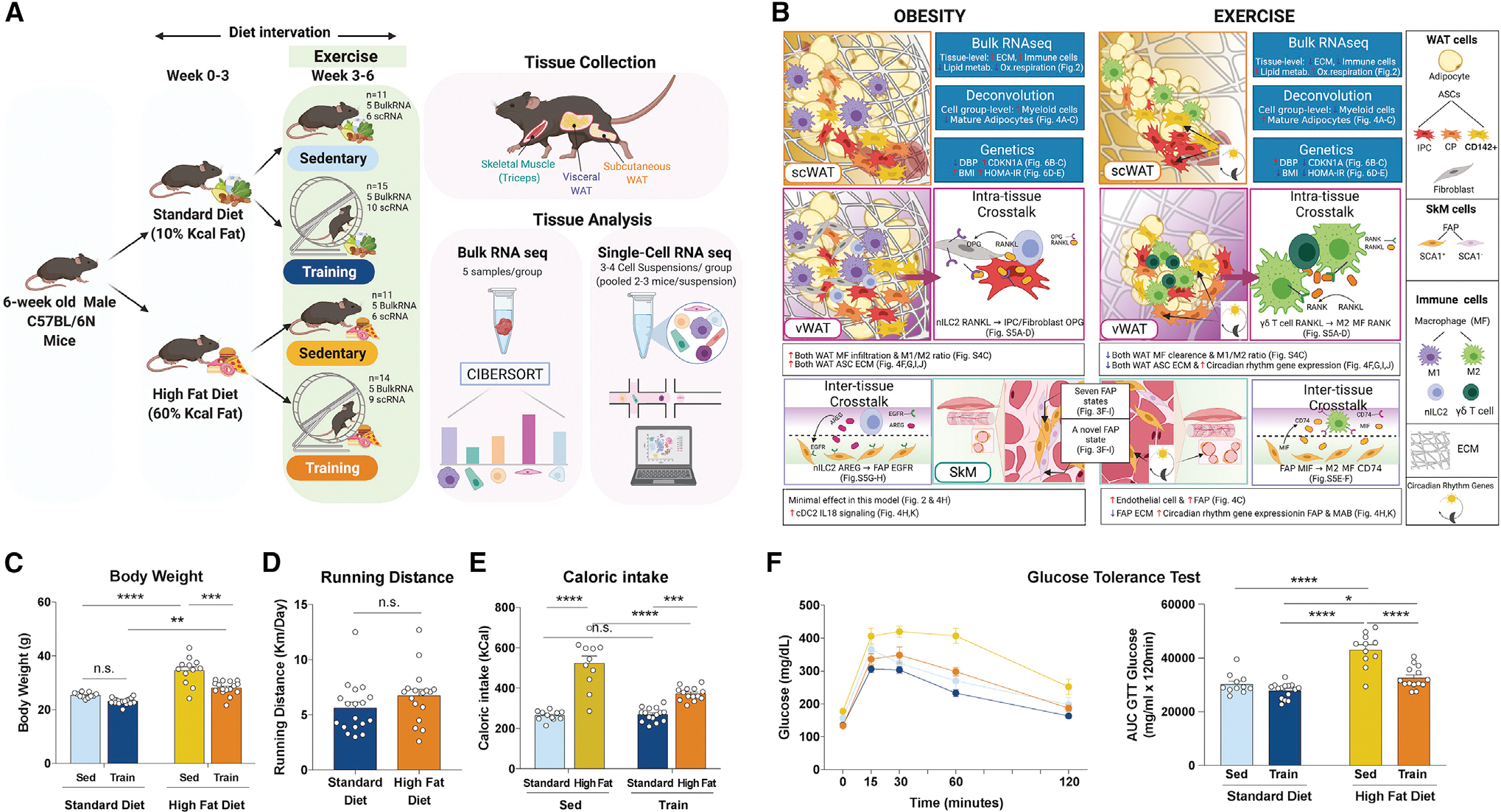
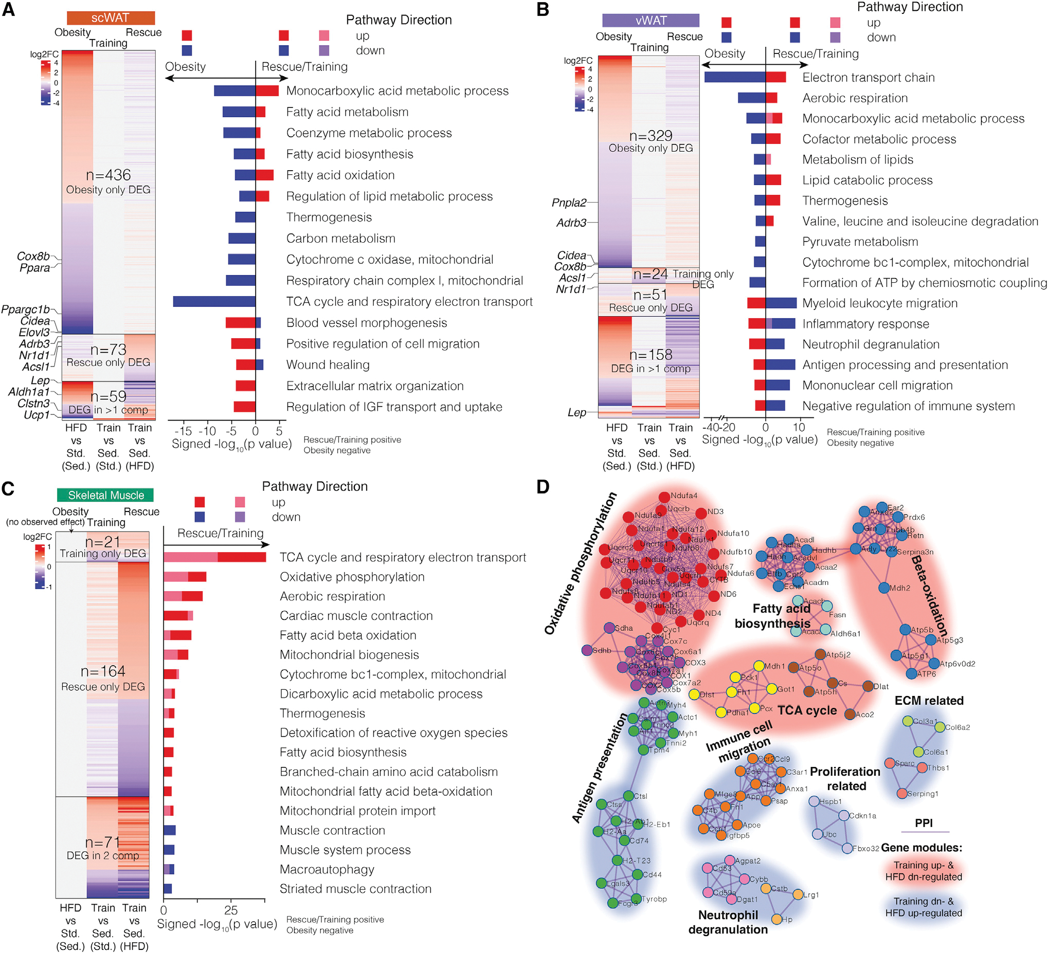
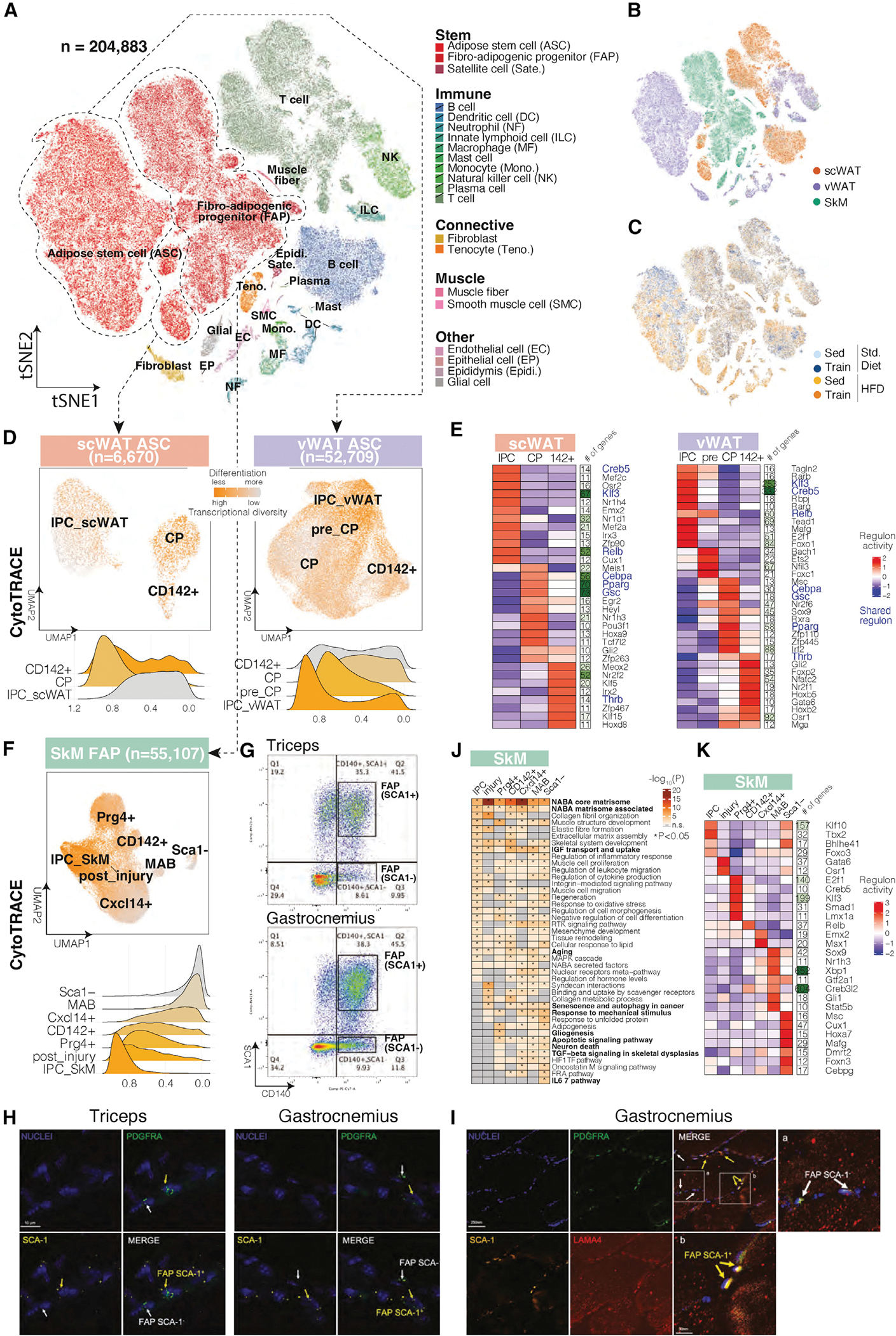
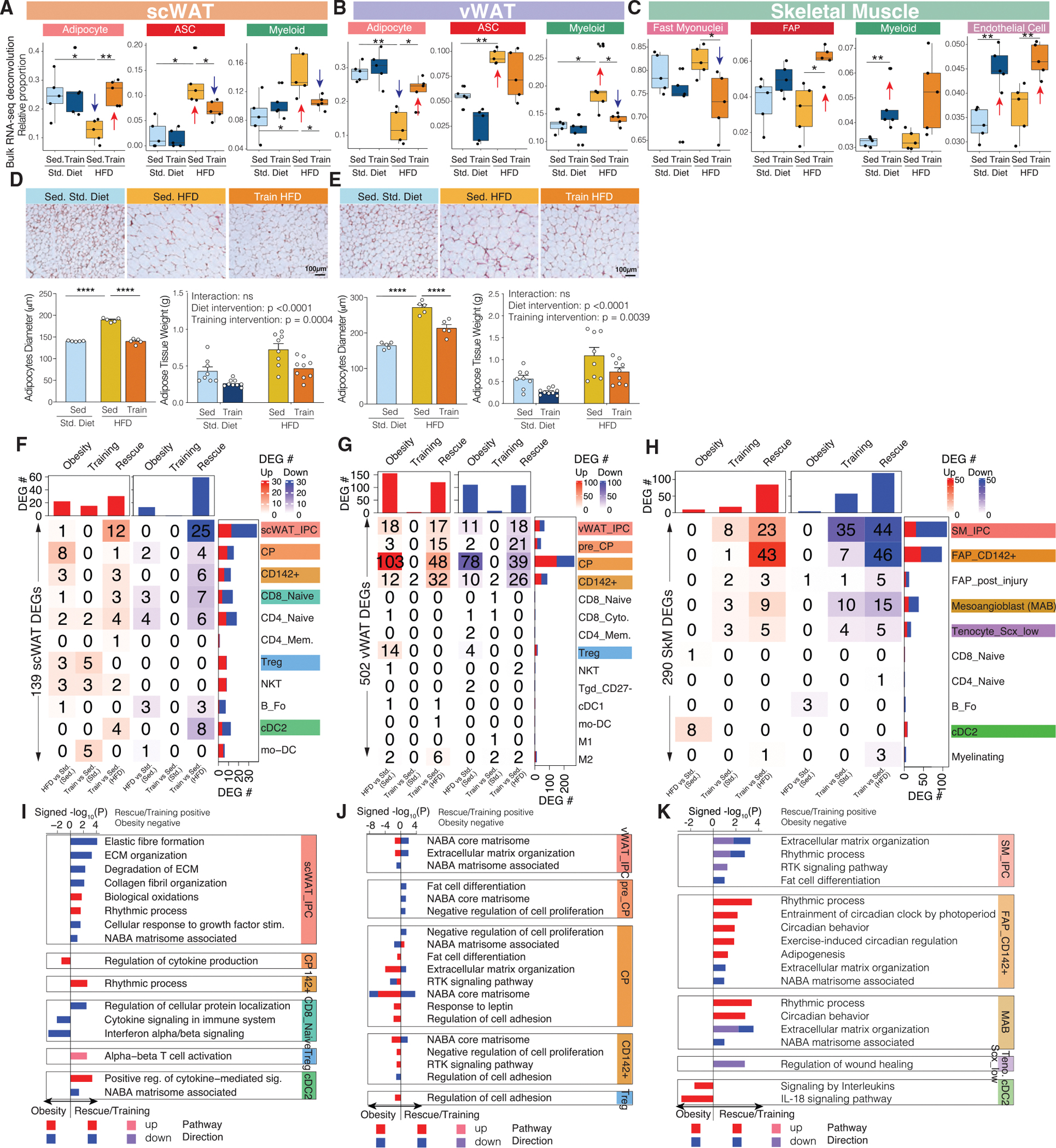
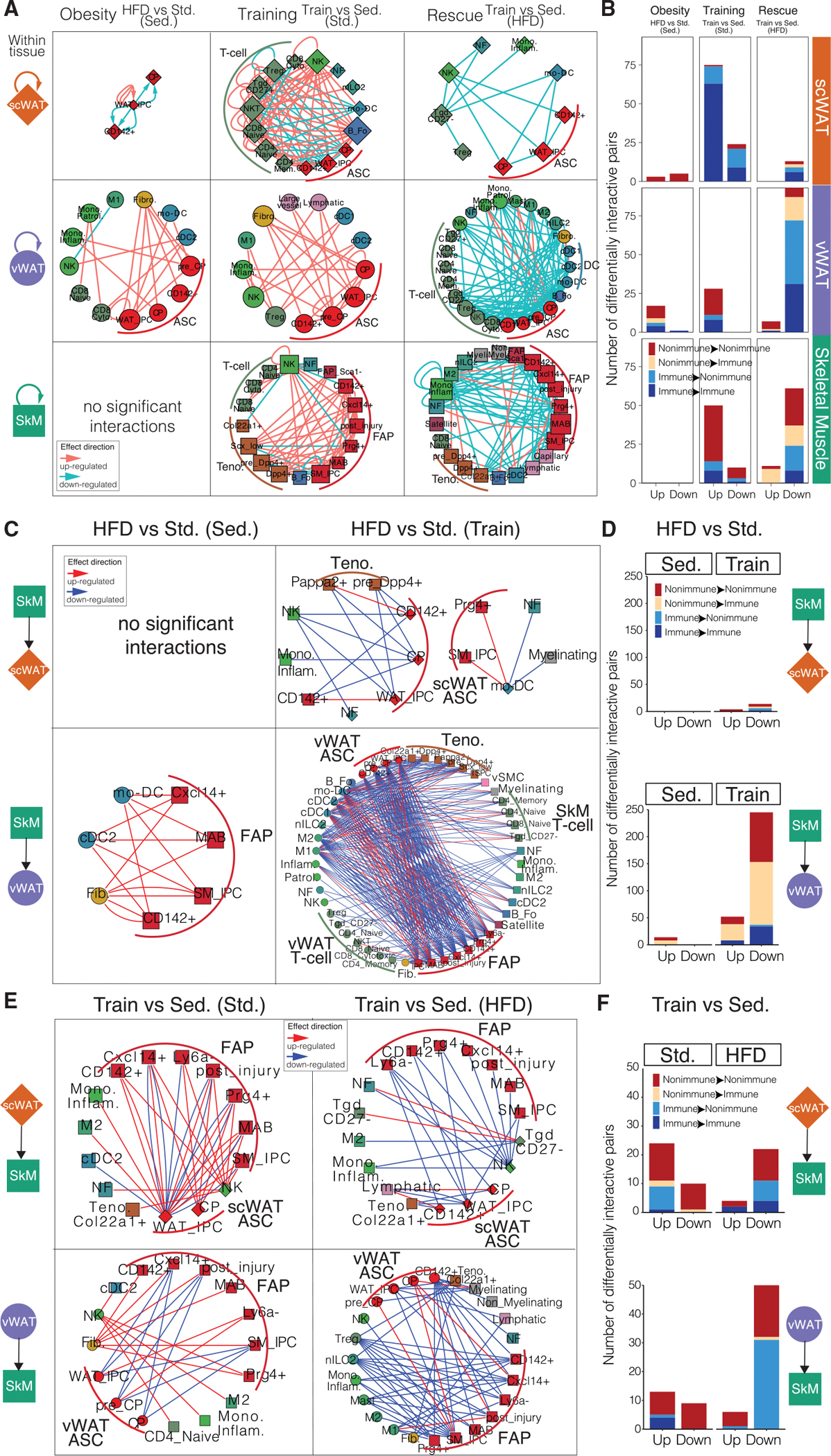
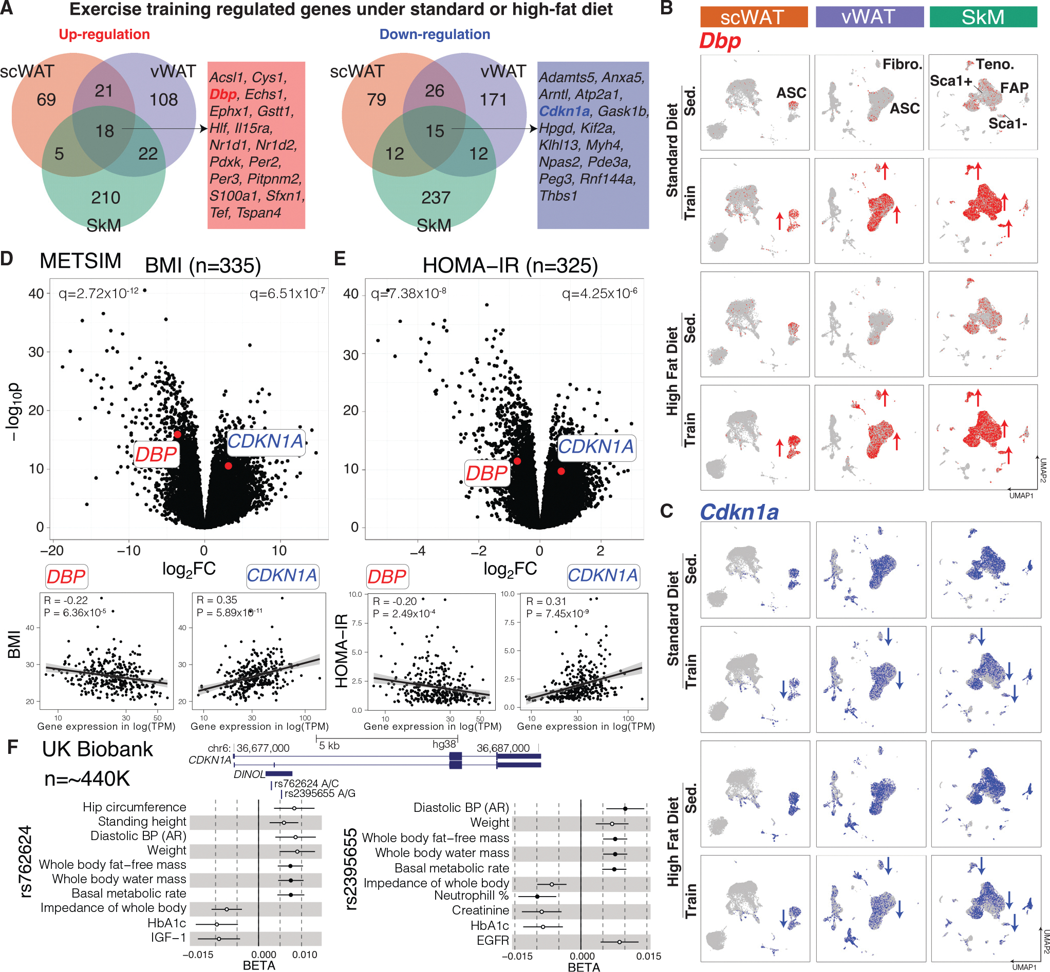
Exercise training is critical for the prevention and treatment of obesity, but its underlying mechanisms remain incompletely understood given the challenge of profiling heterogeneous effects across multiple tissues and cell types. Here, we address this challenge and opposing effects of exercise and high-fat diet (HFD)-induced obesity at single-cell resolution in subcutaneous and visceral white adipose tissue and skeletal muscle in mice with diet and exercise training interventions. We identify a prominent role of mesenchymal stem cells (MSCs) in obesity and exercise-induced tissue adaptation. Among the pathways regulated by exercise and HFD in MSCs across the three tissues, extracellular matrix remodeling and circadian rhythm are the most prominent. Inferred cell-cell interactions implicate within- and multi-tissue crosstalk centered around MSCs. Overall, our work reveals the intricacies and diversity of multi-tissue molecular responses to exercise and obesity and uncovers a previously underappreciated role of MSCs in tissue-specific and multi-tissue beneficial effects of exercise.

Keywords: circadian rhythm pathway; exercise; extracellular matrix remodeling; mesenchymal stem cell; multi-tissue crosstalk; obesity; single-cell atlas; skeletal muscle; white adipose tissue; within-tissue crosstalk.


Conflict of interest statement

Declaration of interests K. Grove is an employee of Novo Nordisk.

Figures

Figure 1.
Figure 1.. Study overview, highlighted results, and phenotypic responses
(A) Overview of the mouse study and tissue profiling. (B) Summary of highlighted results. ECM, extracellular matrix. (C–F) Body weight (C), running distance (D), caloric intake (E), and glucose tolerance test (GTT) result (F) in the four intervention groups. *p < 0.05, **p < 0.01, ***p < 0.001, ****p < 0.0001 by two-way ANOVA followed by Tukey multiple comparison tests. Data are represented as mean ± SEM. AUC, area under curve; n.s., not significant. (A) and (B) were created with BioRender.com. See also Figure S1.
Figure 2.
Figure 2.. Tissue-level transcriptomic responses
(A–C) Genes (heatmap) and pathways (bar plot) that are significantly differentially expressed and enriched across three comparisons: “obesity,” “training,” and “rescue” in scWAT (A), vWAT (B), and SkM (C). The gene heatmap is colored by log2 fold change. The pathway bar plot is colored by pathway direction in the three comparisons (red/pink, upregulated; blue/purple, downregulated). x axis of the bar plot shows −log10 p value with rescue/training pathways being positive and obesity being negative. DEG, differentially expressed gene. (D) Gene networks across selected DEGs from the three tissues that encode interacting proteins, clustered by protein-protein interactions with each cluster named by the most significantly enriched pathway. The cluster is colored by DEG direction with exercise training. sed., sedentary; train, exercise training; std, standard diet. See also Figures S1 and S2 and Table S1.
Figure 3.
Figure 3.. Single-cell atlas and mesenchymal stem cell state characterization
(A) Single-cell atlas of 204,883 cells across three tissues and four intervention groups. The tSNE plot is colored by cell type (warm colors, non-immune cell types; cold colors, immune cell types). (B and C) Single-cell atlas colored by tissue (B) and intervention group (C). (D) Re-clustering of ASCs in scWAT (left) and vWAT (right), colored by CytoTRACE-predicted differentiation stage (orange, less differentiated; gray, more differentiated). Ridge plots of individual ASC states are colored similarly. (E) Clustering of top ASC state-specific regulons (transcription factor with the number of regulated genes as a separate heatmap column) in scWAT (left) and vWAT (right). Shared regulons across the two depots are colored in blue. The heatmap is scaled by column. (F) Re-clustering of FAPs in SkM, colored by CytoTRACE-predicted differentiation stage (orange, less differentiated; gray, more differentiated). Ridge plots of individual FAP states are colored similarly. (G) FACS dot blot showing the sorting gates for Sca1+ and Sca1− FAPs from mouse triceps and gastrocnemius, with the percentages of the two populations labeled. (H) RNA staining of Pdgfra and Ly6a in triceps and gastrocnemius muscle. (I) Immunohistochemistry staining for PDGFRA, SCA-1, and LAMA4 (a marker for muscle fibers) in gastrocnemius muscle. (J and K) Top pathways (J) and regulons (K) enriched in Sca1+ and Sca1− FAPs. The pathway heatmap is colored by −log10 p value. The regulon heatmap is colored by activity score. See also Figure S3 Tables S2 and S3.
Figure 4.
Figure 4.. Single-cell-level proportion and transcriptomic responses across the three tissues
(A–C) Sample-specific proportions of cell types across the four intervention groups in scWAT (A), vWAT (B), and SkM (C) after bulk RNA-seq data deconvolution. The boxplots are defined as Q1 − 1.5*IQR, Q1, median, Q3, and Q3 + 1.5*IQR. (D and E) Histology of scWAT (D) and vWAT (E) across three intervention groups, with bar plots below showing adipocyte diameter and adipose tissue weight changes across intervention groups. (F–H) The number of cell-state-specific DEGs (heatmap) that are upregulated (red) or downregulated (blue) in our three comparisons in scWAT (F), vWAT (G), and SkM (H). (I–K) Pathways (bar plot) that are significantly enriched in cell-state-specific DEGs across the three comparisons in scWAT (I), vWAT (J), and SkM (K). x axis of the bar plot shows −log10 p value with rescue/training pathways being positive and obesity being negative. The bars are colored by pathway direction in the three comparisons (red/pink, upregulated; blue/purple, downregulated). *p < 0.05, **p < 0.01, ***p < 0.001, ****p < 0.0001 by Wilcoxon rank-sum test (A–C) or by two-way ANOVA followed by Tukey multiple comparison tests (D and E). Data are represented as mean ± SEM (D and E). See also Figure S4 Tables S2 and S4.
Figure 5.
Figure 5.. Within- and cross-tissue communication at cell-state level
(A) Within-tissue ligand-receptor networks across the three tissues and three comparisons. Cell states (nodes) are shaped by tissue (diamond, scWAT; circle, vWAT; square, SkM) and sized by outdegree. Ligand-receptor interactions (edges) are directed, from ligand to receptor, and colored by effect direction (pink, upregulated; blue, downregulated). (B) The number of differentially interactive ligand-receptor pairs that are up- and downregulated across the three tissues and three comparisons at the cell-state level. Each bar is colored by whether the ligand or the receptor is from an immune or non-immune cell state. (C and E) Cross-tissue ligand-receptor networks between a pair of tissues and in diet (C) or training (E) comparisons. The nodes and edges are formatted as in (A). (D and F) The number of differentially interactive ligand-receptor pairs that are up- and downregulated between a pair of tissues and in diet (D) or training (F) comparisons at the cell-state level. The bars are colored as in (B). See also Figure S5 Table S5.
Figure 6.
Figure 6.. Two exercise-regulated genes (DBP and CDKN1A) in mice and humans
(A) Overlap of up- and downregulated genes by exercise training under standard or HFD across the three tissues in mice. (B and C) Dbp (B) and CDKN1A (C) expression across the three tissues and four intervention groups. Cell types with the most changes are labeled in the top panel. (D and E) DBP and CDKN1A association with BMI (D) and HOMA-IR (E) in scWAT of METSIM subjects. Genes (dots in upper plots) and subjects (dots in lower plots) are plotted. (F) Association of two SNPs (rs762624 and rs2395655) in CDKN1A with anthropometric and metabolic traits in UK Biobank. The meta-analyzed PheWAS summary statistics (BETAs with standard errors, p < 1–3) are shown. The filled circles are significant after correction (p < 1–5). PheWAS, phenome-wide association study; BP, blood pressure; AR, automated reading. See also Figure S6.

References

    1. Aibar S, González-Blas CB, Moerman T, Huynh-Thu VA, Imrichova H, Hulselmans G, Rambow F, Marine J-C, Geurts P, Aerts J, et al. (2017). SCENIC: single-cell regulatory network inference and clustering. Nat. Methods 14, 1083–1086. - PMC - PubMed
    1. Amezquita RA, Lun ATL, Becht E, Carey VJ, Carpp LN, Geistlinger L, Marini F, Rue-Albrecht K, Risso D, Soneson C, et al. (2020). Orchestrating single-cell analysis with Bioconductor. Nat. Methods 17, 137–145. - PMC - PubMed
    1. Ancel S, Mashinchian O, and Feige JN (2019). Adipogenic progenitors keep muscle stem cells young. Aging 11, 7331–7333. - PMC - PubMed
    1. Aran D, Looney AP, Liu L, Wu E, Fong V, Hsu A, Chak S, Naikawadi RP, Wolters PJ, Abate AR, et al. (2019). Reference-based analysis of lung single-cell sequencing reveals a transitional profibrotic macrophage. Nat. Immunol. 20, 163–172. - PMC - PubMed
    1. Bray NL, Pimentel H, Melsted P, and Pachter L (2016). Erratum: Near-optimal probabilistic RNA-seq quantification. Nat. Biotechnol. 34, 888. - PubMed

Publication types