. 2022 Dec;28(12):1643-1658.
doi: 10.1261/rna.079356.122. Epub 2022 Oct 5.

E. coli 6S RNA complexed to RNA polymerase maintains product RNA synthesis at low cellular ATP levels by initiation with noncanonical initiator nucleotides

Affiliations

E. coli 6S RNA complexed to RNA polymerase maintains product RNA synthesis at low cellular ATP levels by initiation with noncanonical initiator nucleotides

Christopher D Bonar et al. RNA. 2022 Dec.

Abstract

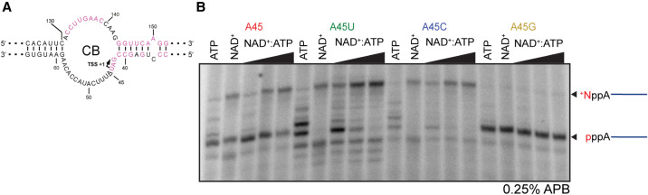
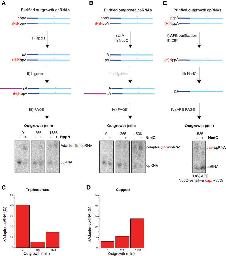
The E. coli 6S RNA is an RNA polymerase (RNAP) inhibitor that competes with σ70-dependent DNA promoters for binding to RNAP holoenzyme (RNAP:σ70). The 6S RNA when bound is then used as a template to synthesize a short product RNA (pRNA; usually 13-nt-long). This pRNA changes the 6S RNA structure, triggering the 6S RNA:pRNA complex to release and allowing DNA-dependent housekeeping gene expression to resume. In high nutrient conditions, 6S RNA turnover is extremely rapid but becomes very slow in low nutrient environments. This leads to a large accumulation of inhibited RNAP:σ70 in stationary phase. As pRNA initiates synthesis with ATP, we and others have proposed that the 6S RNA release rate strongly depends on ATP levels as a proxy for sensing the cellular metabolic state. By purifying endogenous 6S RNA:pRNA complexes using RNA Mango and using reverse transcriptase to generate pRNA-cDNA chimeras, we demonstrate that 6S RNA:pRNA formation can be simultaneous with 6S RNA 5' maturation. More importantly, we find a dramatic accumulation of capped pRNAs during stationary phase. This indicates that ATP levels in stationary phase are low enough for noncanonical initiator nucleotides (NCINs) such as NAD+ and NADH to initiate pRNA synthesis. In vitro, mutation of the conserved 6S RNA template sequence immediately upstream of the pRNA transcriptional start site can increase or decrease the pRNA capping efficiency, suggesting that evolution has tuned the biological 6S RNA sequence for an optimal capping rate. NCIN-initiated pRNA synthesis may therefore be essential for cell viability in low nutrient conditions.

Keywords: 6S RNA; ATP; NAD(H); RNA Mango; exponential phase; pRNA; stationary phase.


Figures

FIGURE 1.
FIGURE 1.
ssrS promoter structure, E. coli 6S RNA secondary structures, pRNA sequence and potential product RNA initiators. (A) Map of the ssrS gene. P1 refers to σ70-dependent Promoter 1, P2 refers to Promoter 2 dependent on both σ38 and σ70 (adapted from Kim 2004, by permission of Oxford University Press). (B) 6S RNA in its free state; resembles a DNA transcriptional bubble. “−35” and “−10” refer to DNA-like regions for RNAP interactions. Highly conserved residues present in the γ-proteobacteria are shown in pink (Shephard et al. 2010). The bubbles to the left (LB1; LB2; LB3) and to the right (RB1; RB2; RB3) of the large central bubble (CB) are noted together with the downstream helix (DH). The pRNA transcription start site (TSS, U44) is indicated by the arrow. (C) Expected 13 nt pRNA sequence for E. coli where X represents p (phosphate) for ATP, +N for NAD+, HN for NADH, or F for FAD initiator incorporation. (D) The 6S RNA:pRNA complex after release from RNAP:σ70 holoenzyme. An intramolecular hairpin (−10:DH hairpin) arises when pRNA elongates by at least 8 nt (Panchapakesan and Unrau 2012); X (shown in red) represents the potential for noncanonical initiation of the release pRNA as shown in panel C. (E) Chemical structures of the known pRNA transcriptional initiator ATP and potential initiators NAD+, NADH, and FAD. The adenosine diphosphate shared by all initiators is indicated in black with the differing functional groups shown in red.
FIGURE 2.
FIGURE 2.
E. coli cells encoding RNA Mango III-tagged 6S RNA accumulate fluorescence during stationary phase. Cells grown overnight in media containing 20 µM TO1-Biotin were imaged at 40× magnification using an EVOS FL Auto 2 microscope. (A) Wild-type (ssrS) and CRISPR-modified (MIII ssrS) cells. Fluorescing cells from staining in TO1-Biotin were identified using the green channel (482 nm excitation and 524 nm emission) (top panels). Cell locations were identified using white light imaging (bottom panels). Scale bar in each panel is 10 µm. (B) Bar graphs quantifying the observed accumulation of green fluorescence in stationary phase cells. Blue refers to the ssrS strain (n = 848), and red refers to the MIII ssrS strain (n = 609). The raw integrated density for each cell was scored using ImageJ. “Range” refers to a set of raw integrated intensities, and “Count” refers to the number of cells quantified for each raw integrated intensity. Histograms were generated using KaleidaGraph.
FIGURE 3.
FIGURE 3.
Mango III-dependent purification of the 6S RNA:pRNA complex and pRNA-cDNA chimera generation. (A) Schematic Mango purification protocol adapted from Panchapakesan et al. (2017) to include pRNA-cDNA chimera (cpRNA) synthesis. (I) Crude MIII ssrS RNA extracts were prepared from chilled, methanol fixed cell culture samples. 6S RNAM is shown in black with conserved regions in pink, and the MIII tag in orange. Hybridized pRNA in a 6S RNAM:pRNA complex is shown in blue. Extraneous cellular RNA is shown in brown. (II) 6S RNAM:pRNA complexes were immobilized on streptavidin magnetic beads (“S”) derivatized with TO1-Biotin (shown in green and light gray) and washed. (III) Reverse transcription (RT) generates bead-immobilized pRNA–DNA (cpRNA) chimeras. The cDNA of cpRNA is shown in light blue. (IV) Formamide elution recovers cpRNAs for further downstream analysis. (B) Secondary structure of a 6S RNAM:cpRNA complex bound to streptavidin beads. Radioactive cpRNA was generated by spiking the RT step with α-32P dATP. “B” of TO1-Biotin refers to biotin bound to streptavidin, and “TO1” refers to thiazole orange. For fully matured 6S RNA, this cpRNA is 44-nt-long. Dashed lines at the 6S RNAM 5′ end (cpRNA 3′ end) imply potentially longer 6S RNA and cDNA lengths resulting from immature 6S RNA.
FIGURE 4.
FIGURE 4.
A range of 6S RNA processing events are associated with pRNA production in early outgrowth time points. (A) RNA transcription and ribonucleases process the 5′ termini of the 6S RNA (Kim 2004) prior to or during loading into the inhibitory complex, which are then released by pRNA synthesis. The RNAP core is shown in teal, and σ70 is shown in blue. (B) cpRNAs as a function of outgrowth time. Each outgrowth cpRNA sample purified from MIII ssrS bacteria was loaded either without or with alkaline hydrolysis treatment so as to measure the cDNA size. Dideoxy size ladders were generated by RT extension of synthetic DNA primer 5′-ATC GGC TCA GGG G-3′ adapted from the expected pRNA sequence. cpRNAs were labeled with α-32P dATP using RT. (C) Individual bands found in the 4 min time point shown in panel B were excised and subjected to complete alkaline hydrolysis. Bands are ordered by decreasing cpRNA length from left to right. The cpRNA bands indicative of 5′-precursor and matured 6S RNA species are marked by bold length numbers in the gap between the two gels. All samples purified by Mango and PAGE were normalized by cpRNA concentration and resolved using 10% denaturing PAGE. (D) Observed cpRNA sizes are related to 5′ mature 6S RNA as well as primary transcripts initiated at promoters P2 and P1.
FIGURE 5.
FIGURE 5.
6S RNA release is enabled in vitro by NCINs when ATP is absent. Native shift assay of internally radiolabeled synthetic 6S RNAM releasing from RNAP, relative to free 6S RNAM and 6S RNAM:RNAP:σ70 controls (”Free” and “Bound”). IN refers to each “initiator nucleotide”: ATP, NAD+, NADH, and FAD. Two negative controls lacking ATP and NCIN were prepared: “free” (6S RNAM) and “bound” (6S RNAM:RNAP:σ70 complex). ATP was then added at 150 µM and 1000 µM as two positive controls. Each NCIN reaction was performed in the absence of ATP (“−ATP“) for 32 min using the indicated concentrations of NCIN. Samples were normalized by 6S RNAM specific activity and resolved in a 5% native gel containing 5% glycerol at 4°C.
FIGURE 6.
FIGURE 6.
NAD(H) and FAD-modified pRNA products produced in vitro. (A) Denaturing gel analysis of NAD(H) containing pRNAs produced in vitro. (B) APB denaturing gel analysis of the same samples. Both gels in panels A and B were resolved using 20% denaturing PAGE. (C) RNA processing by CIP, RppH and NudC. The shared adenosine diphosphate is shown in black, whereas the differing upstream moiety is shown in red. X refers to a 5′ cap. (D) 5′ processing of in vitro synthesized pRNA using γ-32P ATP and processed with CIP and RppH. (E) 5′ processing of in vitro synthesized α-32P UTP radiolabeled pRNA species using CIP, RppH, and NudC. “(pp)A” refers to the 5′ terminal end of pRNAs containing either “ppA” or “A” (5′-OH), as these pRNA species migrate with similar velocities. 5′-triphosphorylated (“pppA”) pRNAs (ATP ± CIP conditions) shown in panel E run faster than A-pRNAs, given that pppA–pRNAs have more negative charges. The pA–pRNAs migrate faster than the pppA–pRNAs (panel E, lanes ATP ± RppH) due to less weight. These band patterns reflect a mixture of pRNAs initiated with a capped adenosine (slower mobility; NAD[H] or FAD) or ATP (fast mobility). Low amounts of HNppA–pRNA (NADH–pRNA) band are likely due to 5′ oxidation, resulting in +NppA–pRNA (NAD+-pRNA). The ppA–pRNAs are assumed to have come from cap degradation. Samples in panels D and E were normalized to the same pRNA specific activity after Mango purification and resolved using 23% denaturing PAGE containing 0.25% APB.
FIGURE 7.
FIGURE 7.
NAD+ capping efficiency is strongly modulated by 6S RNA sequence in vitro. (A) 6S RNA with A45 underlined. (B) Competitive in vitro pRNA synthesis using A45N 6S RNA templates with NAD+ concentration held constant at 1 mM and ATP decreasing from 100 to 10 to 1 µM. Samples were normalized by pRNA specific activity and resolved using 23% denaturing PAGE containing 0.25% APB.
FIGURE 8.
FIGURE 8.
In vivo NCINs are used for pRNA initiation as cells enter stationary phase. Preparation and diagnosis of the 5′ end of Mango-purified, PAGE-purified outgrowth cpRNAs radiolabeled with α-32P dATP. (A) Triphosphate cpRNA ligation procedure. (I) RppH treatment. (II) 5′ RNA adapter (purple) ligation. (III) Separation of adapter ligation products and unligated cpRNA by 8% denaturing PAGE. (B) Capped cpRNA ligation procedure. (I) CIP treatment. (II) NudC treatment. Both ligation (III) and PAGE (IV) steps are the same as in II and III of panel A. (C) Quantification of ligation percentage from panel A triphosphate cpRNA procedure. (D) Quantification of ligation percentage from panel B capped cpRNA procedure. (E) Capped cpRNA APB gel analysis (1536 min of outgrowth, only). (I) APB Purification using 10% denaturing PAGE containing 0.8% APB. (II) CIP treatment. (III) NudC treatment. (IV) 10% denaturing PAGE containing 0.8% APB to resolve the cpRNAs. The histograms in panels C and D are determined from a single experiment.
Christopher D. Bonar

References

    1. Autour A, Jeng S CY, Cawte A D, Abdolahzadeh A, Galli A, Panchapakesan SSS, Rueda D, Ryckelynck M, Unrau PJ. 2018. Fluorogenic RNA Mango aptamers for imaging small non-coding RNAs in mammalian cells. Nat Commun 9: 656. 10.1038/s41467-018-02993-8 - DOI - PMC - PubMed
    1. Barrick JE, Sudarsan N, Weinberg Z, Ruzzo WL, Breaker RR. 2005. 6S RNA is a widespread regulator of eubacterial RNA polymerase that resembles an open promoter. RNA 11: 774–784. 10.1261/rna.7286705 - DOI - PMC - PubMed
    1. Beckmann BM, Hoch PG, Marz M, Willkomm DK, Salas M, Hartmann RK. 2012. A pRNA-induced structural rearrangement triggers 6S-1 RNA release from RNA polymerase in Bacillus subtilis: pRNA length-dependent change in 6S-1 RNA structure. EMBO J 31: 1727–1738. 10.1038/emboj.2012.23 - DOI - PMC - PubMed
    1. Bennett BD, Kimball EH, Gao M, Osterhout R, Van Dien SJ, Rabinowitz JD. 2009. Absolute metabolite concentrations and implied enzyme active site occupancy in Escherichia coli. Nat Chem Biol 5: 593–599. 10.1038/nchembio.186 - DOI - PMC - PubMed
    1. Bird JG, Zhang Y, Tian Y, Panova N, Barvík I, Greene L, Liu M, Buckley B, Krásnỳ L, Lee JK. 2016. The mechanism of RNA 5′ capping with NAD+, NADH and desphospho-CoA. Nature 535: 444–447. 10.1038/nature18622 - DOI - PMC - PubMed

Publication types

MeSH terms

LinkOut - more resources