

Case Reports
doi: 10.2169/internalmedicine.0489-22.
Epub 2022 Oct 5.
Yersinia enterocolitica Enteritis Diagnosed with Erythema Nodosum
Affiliations
- PMID: 36198596
- PMCID: PMC10258089
- DOI: 10.2169/internalmedicine.0489-22
Item in Clipboard
Case Reports
Yersinia enterocolitica Enteritis Diagnosed with Erythema Nodosum
Intern Med.
.
Abstract
We herein report a rare case of Yersinia enterocolitica enteritis with a fever and abdominal pain followed by erythema nodosum (EN) a few days later. The diagnosis was confirmed based on characteristic colonoscopy and computed tomography findings, pathology, and mucosal culture. Yersinia enteritis is a curable disease provided a proper diagnosis and treatment are performed. Although EN is a rare clinical course, it should still be considered as a differential diagnosis.
Keywords: Yersinia enterocolitica enteritis; erythema nodosum.
Conflict of interest statement
Figures

Skin findings on the lower leg. a, b: Multiple tender infiltrative erythematous plaques are visible on the extensor surfaces of both lower legs (red arrows).

Computed tomography. a: Swollen lymph nodes are noted around the ileocecal area (red arrow). b: Wall thickening of the ileocecal region is evident (red arrow).

Colonoscopy. a: Ileum end with WLI: Scattered aphthous ulcers and erosion. b: Ileum end with WLI. c: Ileum end with indigo carmine dyeing: Multiple round ulcers. d: Cecum with WLI. e: Cecum-ascending colon with WLI. f: Ascending colon with WLI: Small erosions scattered in the cecum, ileocecal valve, and ascending colon. WLI: white-light imaging

Pathological findings of the ileum end and cecum. a: Ileum end [Hematoxylin and Eosin (H&E) staining×40]: A collection of lymphocytes can be seen in the submucosal layer. b: Ileum end (H&E staining×100): Infiltration of lymphocytes and a small number of neutrophils can be seen in the mucosal intrinsic layer. b: Cecum (H&E staining×100): A collection of lymphocytes is seen in the mucosal and submucosal layers. No germinal center or granuloma formation is seen.
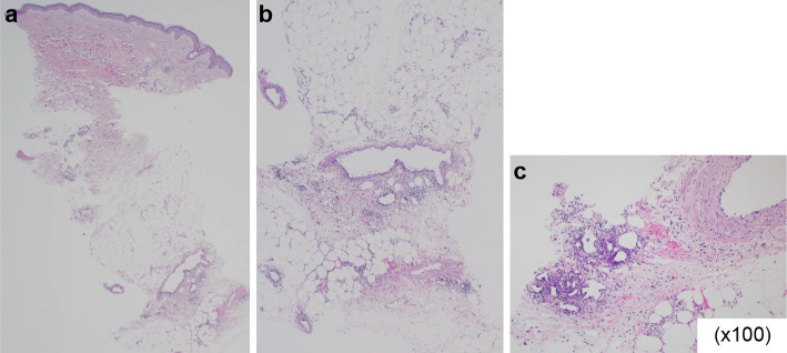
Pathological findings of erythematous area of rash. a: A skin biopsy [Hematoxylin and Eosin (H&E) staining×20]. b: (H&E staining×40): Inflammatory cell infiltration and fibrosis are most evident in the fat septum in the subcutaneous fatty tissue. c: A skin biopsy (H&E staining×100): Lymphocytic infiltration, fibrosis, and septal panniculitis in the fat septum of the subcutaneous fatty tissue.

Skin findings on the lower leg after two weeks. a, b: Erythema on the extensor surfaces of both lower legs resolved (red arrows).

Clinical course of Yersiniaenterocolitica enteritis with erythema nodosum. Abdominal pain was observed 6 days before the onset of erythema nodosum. Levofloxacin Hydrate administration rapidly improved the symptoms, and EN was cured in approximately two weeks.

Colonoscopy findings after one year. a: Ileum end. b: Cecum. c: Cecum-ascending colon. d: Ascending colon: Erosions and ulcers in the ileocecal area have improved.
References
-
- Cover TL, Aber RC. Yersinia enterocolitica. N Engl J Med 321: 16-24, 1989. - PubMed
-
- Vantrappen G, Ponette E, Geboes K, Bertrand P. Yersinia enteritis and enterocolitis: gastroenterological aspects. Gastroenterology 72: 220-227, 1977. - PubMed
-
- Winblad S. Erythema nodosum associated with infection with Yersinia enterocolitica. Scand J Infect Dis 1: 11-16, 1969. - PubMed
-
- Cribier B, Caille A, Heid E, Grosshans E. Erythema nodosum and associated diseases. A study of 129 cases. Int J Dermatol 37: 667-672, 1998. - PubMed
-
- Leung AKC, Leong KF, Lam JM. Erythema nodosum. World J Pediatr 14: 548-554, 2018. - PubMed
Publication types
MeSH terms
LinkOut - more resources
Full Text Sources
Research Materials