Review
. 2023 Mar;20(3):197-210.
doi: 10.1038/s41569-022-00770-1. Epub 2022 Oct 5.

Vascular endothelial cell development and diversity

Affiliations
Review

Vascular endothelial cell development and diversity

Emily Trimm et al. Nat Rev Cardiol. 2023 Mar.

Abstract

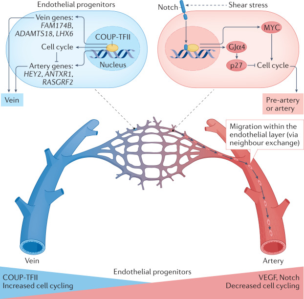
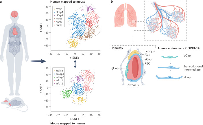
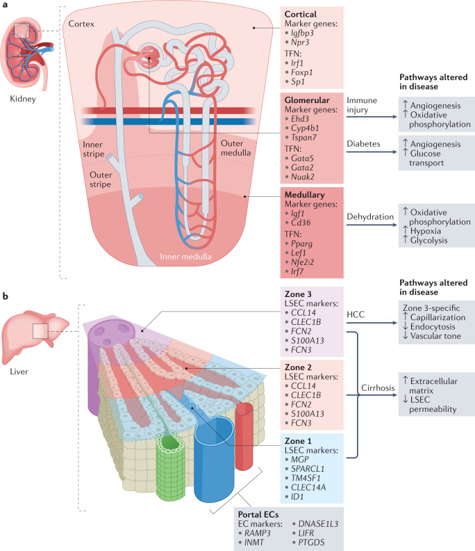
Vascular endothelial cells form the inner layer of blood vessels where they have a key role in the development and maintenance of the functional circulatory system and provide paracrine support to surrounding non-vascular cells. Technical advances in the past 5 years in single-cell genomics and in in vivo genetic labelling have facilitated greater insights into endothelial cell development, plasticity and heterogeneity. These advances have also contributed to a new understanding of the timing of endothelial cell subtype differentiation and its relationship to the cell cycle. Identification of novel tissue-specific gene expression patterns in endothelial cells has led to the discovery of crucial signalling pathways and new interactions with other cell types that have key roles in both tissue maintenance and disease pathology. In this Review, we describe the latest findings in vascular endothelial cell development and diversity, which are often supported by large-scale, single-cell studies, and discuss the implications of these findings for vascular medicine. In addition, we highlight how techniques such as single-cell multimodal omics, which have become increasingly sophisticated over the past 2 years, are being utilized to study normal vascular physiology as well as functional perturbations in disease.


Conflict of interest statement

The authors declare no competing interests.

Figures

Fig. 1
Fig. 1. Artery and vein endothelial cell differentiation in a developing vascular bed.
Artery differentiation is coupled to cell cycle arrest. Shear stress activation of Notch signalling upregulates the levels of gap junction α4 protein (GJα4), which subsequently inhibits the expression of cell cycle genes via p27 expression. Notch signalling also inhibits MYC-induced transcription of genes related to cell cycle and metabolism. Together, Notch signalling indirectly supports arterial cell fate via cell cycle inhibition. Pre-artery endothelial cells then migrate against the direction of blood flow in the vein-to-artery direction. Adjacent endothelial cells exchange positions in a process known as ‘neighbour exchange’ to expand arteries. By contrast, COUP transcription factor 2 (COUP-TFII) (also known as NR2F2) activates the expression of cell cycle genes, inhibits the expression of certain genes associated with arterial specification and induces vein-specific gene expression. VEGF, vascular endothelial growth factor.
Fig. 2
Fig. 2. scRNA-seq reveals novel endothelial cell types.
a | Comparison of human and mouse single-cell RNA sequencing (scRNA-seq) datasets for heart and brain vasculature reveals similar endothelial cell states between species. Although expression of unique genes exists for endothelial cell subtypes within each species, these analyses support the use of mouse models for studying human development and pathology. Key findings from these datasets reveal that the heart has conserved endothelial cell populations and similar capillary endothelial cell states in mice and humans and unique human artery specification genes. Furthermore, the brain also shows conserved endothelial cell populations and zonation between mice and humans and a greater diversity of human perivascular cells. b | Two transcriptionally distinct populations of capillary endothelial cells exist in the lung: general capillaries (gCap) and aerocyte capillaries (aCap). These two capillary populations have different roles in lung maturation and mature lung function. In disease states such as adenocarcinoma, a transcriptional intermediate between gCap and aCap arises in both humans and mice. This transcriptional intermediate has also been seen in the lungs of patients with coronavirus disease 2019 (COVID-19). Overlapping features of inflammation and cellular stress can also be seen in both capillary subtypes in advanced chronic obstructive pulmonary disease. AV1, alveolar type 1 cell; RBC, red blood cell; t-SNE, t-distributed stochastic neighbour embedding.
Fig. 3
Fig. 3. scRNA-seq reveals inter-organ heterogeneity and heterogeneous endothelial cell responses to disease.
a | Single-cell RNA sequencing (scRNA-seq) has revealed transcriptomic heterogeneity between endothelial cells in each compartment of the kidney. Cortical endothelial cells, glomerular endothelial cells and medullary endothelial cells have differential responses to injury. Marker genes and enriched transcriptional factor networks (TFN) for each subtype are indicated. Mouse models have been developed to assess kidney injury during disease states including immune-mediated injury (nephritis and systemic lupus erythematosus), diabetic kidney injury and dehydration. Key pathways that are altered in endothelial cells from the different renal compartments of mice are highlighted. b | Endothelial cell zonation in the liver has been characterized in both humans and mice. Broadly, liver sinusoid endothelial cells (LSECs) can be grouped into three zones based on their localization with respect to the portal vein and central vein. The top five differentially expressed genes for human LSECs are indicated as marker genes for each zone. Studies in mice have revealed that LSECs in zone 3 are the most susceptible to damage associated with cirrhosis. EC, endothelial cell; HCC, hepatocellular carcinoma.

References

    1. World Health Organization. The Top 10 Causes of Death (WHO, 2020).
    1. Paik DT, et al. Single-cell RNA sequencing unveils unique transcriptomic signatures of organ-specific endothelial cells. Circulation. 2020;142:1848–1862. doi: 10.1161/CIRCULATIONAHA.119.041433. - DOI - PMC - PubMed
    1. Elmentaite R, Domínguez Conde C, Yang L, Teichmann SA. Single-cell atlases: shared and tissue-specific cell types across human organs. Nat. Rev. Genet. 2022 doi: 10.1038/s41576-022-00449-w. - DOI - PubMed
    1. den Braanker H, van Stigt AC, Kok MR, Lubberts E, Bisoendial RJ. Single-cell RNA sequencing reveals heterogeneity and functional diversity of lymphatic endothelial cells. Int. J. Mol. Sci. 2021 doi: 10.3390/ijms222111976. - DOI - PMC - PubMed
    1. Corada M, Morini MF, Dejana E. Signaling pathways in the specification of arteries and veins. Arterioscler. Thromb. Vasc. Biol. 2014;34:2372–2377. doi: 10.1161/ATVBAHA.114.303218. - DOI - PubMed

Publication types