

Alzheimer's disease-related transcriptional sex differences in myeloid cells
- PMID: 36199077
- PMCID: PMC9535846
- DOI: 10.1186/s12974-022-02604-w
Alzheimer's disease-related transcriptional sex differences in myeloid cells
Abstract
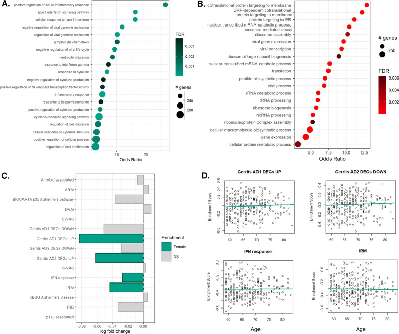
Sex differences have been identified in many diseases associated with dysregulated immune responses, including Alzheimer's disease (AD), for which approximately two-thirds of patients are women. An accumulating body of research indicates that microglia may play a causal role in the pathogenesis of this disease. We hypothesised that sex differences in the transcriptome of human myeloid cells may contribute to the sex difference observed in AD prevalence. To explore this, we assessed bulk and single-nuclear RNA sequencing data sets generated from four human derived myeloid cell populations: post-mortem microglial nuclei, peripheral monocytes, monocyte-derived macrophages (MDMs) and induced pluripotent stem cell derived microglial-like cells (MGLs). We found that expression of AD risk genes, gene signatures associated with the inflammatory response in AD, and genes related to proinflammatory immune responses were enriched in microglial nuclei isolated from aged female donors without ante-mortem neurological disease, relative to those from males. In addition, these inflammation-associated gene sets were found to be enriched in peripheral monocytes isolated from postmenopausal women and in MDMs obtained from premenopausal individuals relative to age-matched males. Expression of these gene sets did not differ in MDMs derived from women whose blood was sampled across the menstrual cycle or in MGLs cultured with 17β-oestradiol. This suggests that the observed gene set enrichments in myeloid cells from women were not being driven by acute hormonal influences. Together, these data support the hypothesis that the increased prevalence of AD in women may be partly explained by a myeloid cell phenotype biased towards expression of biological processes relevant to AD.
Keywords: Microglia; Neurodegeneration; Neuroinflammation; Sex.
© 2022. The Author(s).
Conflict of interest statement
PMM has received consultancy fees from Novartis and Biogen. He has received honoraria or speakers’ fees from Novartis and Biogen and has received research or educational funds from Biogen, Novartis, Merck and Bristol Myers Squibb.
Figures


Similar articles
-
Microglial senescence contributes to female-biased neuroinflammation in the aging mouse hippocampus: implications for Alzheimer's disease.J Neuroinflammation. 2023 Aug 16;20(1):188. doi: 10.1186/s12974-023-02870-2. J Neuroinflammation. 2023. PMID: 37587511 Free PMC article.
-
Myeloid Arginase 1 Insufficiency Exacerbates Amyloid-β Associated Neurodegenerative Pathways and Glial Signatures in a Mouse Model of Alzheimer's Disease: A Targeted Transcriptome Analysis.Front Immunol. 2021 May 11;12:628156. doi: 10.3389/fimmu.2021.628156. eCollection 2021. Front Immunol. 2021. PMID: 34046031 Free PMC article.
-
CD11a expression distinguishes infiltrating myeloid cells from plaque-associated microglia in Alzheimer's disease.Glia. 2019 May;67(5):844-856. doi: 10.1002/glia.23575. Epub 2018 Dec 26. Glia. 2019. PMID: 30588668 Free PMC article.
-
Microglia heterogeneity and neurodegeneration: The emerging paradigm of the role of immunity in Alzheimer's disease.J Neuroimmunol. 2020 Apr 15;341:577185. doi: 10.1016/j.jneuroim.2020.577185. Epub 2020 Feb 3. J Neuroimmunol. 2020. PMID: 32045774 Review.
-
The interplay between microglial states and major risk factors in Alzheimer's disease through the eyes of single-cell RNA-sequencing: beyond black and white.J Neurophysiol. 2019 Oct 1;122(4):1291-1296. doi: 10.1152/jn.00395.2019. Epub 2019 Jul 31. J Neurophysiol. 2019. PMID: 31365320 Review.
Cited by
-
Single cell transcriptome analysis of the THY-Tau22 mouse model of Alzheimer's disease reveals sex-dependent dysregulations.Cell Death Discov. 2024 Mar 7;10(1):119. doi: 10.1038/s41420-024-01885-9. Cell Death Discov. 2024. PMID: 38453894 Free PMC article.
-
Sex differences and the role of estrogens in the immunological underpinnings of Alzheimer's disease.Alzheimers Dement (N Y). 2025 Aug 17;11(3):e70139. doi: 10.1002/trc2.70139. eCollection 2025 Jul-Sep. Alzheimers Dement (N Y). 2025. PMID: 40827126 Free PMC article. Review.
-
Tlr7 drives sex differences in age- and Alzheimer's disease-related demyelination.Science. 2024 Nov 29;386(6725):eadk7844. doi: 10.1126/science.adk7844. Epub 2024 Nov 29. Science. 2024. PMID: 39607927 Free PMC article.
-
Genetic and Epigenetic Sexual Dimorphism of Brain Cells during Aging.Brain Sci. 2023 Jan 24;13(2):195. doi: 10.3390/brainsci13020195. Brain Sci. 2023. PMID: 36831738 Free PMC article. Review.
-
Relationship between sex biases in gene expression and sex biases in autism and Alzheimer's disease.medRxiv [Preprint]. 2023 Sep 1:2023.08.29.23294773. doi: 10.1101/2023.08.29.23294773. medRxiv. 2023. Update in: Biol Sex Differ. 2024 Jun 7;15(1):47. doi: 10.1186/s13293-024-00622-2. PMID: 37693465 Free PMC article. Updated. Preprint.
References
MeSH terms
Substances
Grants and funding
LinkOut - more resources
Full Text Sources
Medical
