

Cutaneous metastasis of carcinomatous component of ovarian carcinosarcoma: A case report and review of the literature
- PMID: 36199118
- PMCID: PMC9533490
- DOI: 10.1186/s13000-022-01256-x
Cutaneous metastasis of carcinomatous component of ovarian carcinosarcoma: A case report and review of the literature
Abstract
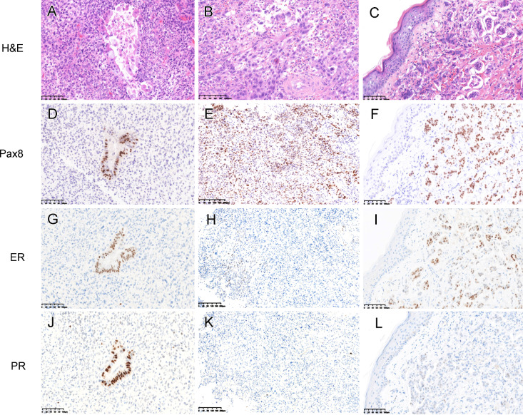
Skin metastasis of ovarian cancer is extremely rare. We report an unusual case of ovarian carcinosarcoma with cutaneous metastasis of carcinomatous component that displayed distinct clinical manifestation. A 48-year-old woman presented to the dermatologist complaining of a new onset of erythematous, plaque-like skin rash with multiple small nodules on the left inner thigh, the area measuring 8 × 5cm. While the patient had no history of dermatologic conditions, she underwent a total hysterectomy and bilateral salpingo-oophorectomy, omentectomy, and lymph node dissection 16 months ago with a pathology confirmed stage IIIC ovarian carcinosarcoma. Of note, the carcinomatous component, mainly adenocarcinoma with hybrid features of seromucinous, endometrioid and minor high-grade serous carcinoma, involved bilateral fallopian tubes, omentum, and parametrium with extensive lymph node metastases. A skin biopsy specimen revealed an adenocarcinoma involving epidermis, dermis, and subcutaneous tissue with nodular contours, consistent with metastatic carcinomatous component of carcinosarcoma. Both carcinomatous component of primary ovarian carcinosarcoma and metastatic adenocarcinoma in the skin demonstrated Pax8, WT-1, and ER positivity and a mutation pattern of p53. The patient passed away 15 months after identification of skin metastasis. This case represents a unique example of cutaneous metastasis of ovarian carcinosarcoma with distinct clinical manifestation and detailed histopathological description. Alertness to the possibility of cutaneous metastasis, in combination with clinical history, morphological and immunohistochemical findings, is critical for a definitive classification.
Keywords: Cutaneous metastasis; Malignant mixed müllerian tumor; Ovarian carcinosarcoma.
© 2022. The Author(s).
Conflict of interest statement
The authors have disclosed that they have no significant relationships with, or financial interest in, any commercial companies pertaining to this article.
Figures


Similar articles
-
A rare case of ovarian carcinosarcoma with squamous cell carcinoma.J Ovarian Res. 2019 Apr 4;12(1):32. doi: 10.1186/s13048-019-0507-3. J Ovarian Res. 2019. PMID: 30947745 Free PMC article.
-
Carcinosarcoma arising from high-grade serous carcinoma of the ovary without estrogen receptor, WT-1, and PAX8 immunoreactivity.Taiwan J Obstet Gynecol. 2022 Jan;61(1):110-114. doi: 10.1016/j.tjog.2021.10.001. Taiwan J Obstet Gynecol. 2022. PMID: 35181017
-
Folliculotropic Cutaneous Metastases and Lymphangitis Carcinomatosa: When Cutaneous Metastases of Breast Carcinoma Are Mistaken for Cutaneous Infections.Acta Dermatovenerol Croat. 2016 Jun;24(2):154-7. Acta Dermatovenerol Croat. 2016. PMID: 27477179
-
[Carcinosarcoma of the skin].Ann Dermatol Venereol. 2006 Apr;133(4):362-5. doi: 10.1016/s0151-9638(06)70916-4. Ann Dermatol Venereol. 2006. PMID: 16733452 Review. French.
-
Unusual Cutaneous Metastasis of Uterine Carcinosarcoma: A Case Report and Review of the Literature.Am J Dermatopathol. 2016 May;38(5):366-9. doi: 10.1097/DAD.0000000000000476. Am J Dermatopathol. 2016. PMID: 26675357 Review.
Cited by
-
Cutaneous Ovarian Carcinoma Metastases: Case Report and Literature Review.Cureus. 2023 Aug 31;15(8):e44459. doi: 10.7759/cureus.44459. eCollection 2023 Aug. Cureus. 2023. PMID: 37791206 Free PMC article.
-
Cutaneous Metastases-Histological Particularities of Multifaceted Entities.Dermatopathology (Basel). 2025 Apr 25;12(2):14. doi: 10.3390/dermatopathology12020014. Dermatopathology (Basel). 2025. PMID: 40407482 Free PMC article.
-
Bilateral ovarian carcinosarcoma - A rare and aggressive malignancy: Case report.Int J Surg Case Rep. 2025 Mar;128:111066. doi: 10.1016/j.ijscr.2025.111066. Epub 2025 Feb 15. Int J Surg Case Rep. 2025. PMID: 39961175 Free PMC article.
References
-
- Kurman RJ, Carcangiu ML, Herrington CS, et al. WHO Classification of Tumors of Female Reproductive Organs. Lyon: IARC; 2014.
-
- Fujii H, Yoshida M, Gong ZX, et al. Frequent genetic heterogeneity in the clonal evolution of gynecological carcinosarcoma and its influence on phenotypic diversity. Cancer Res Jan. 2000;1:114–20. - PubMed
Publication types
MeSH terms
Substances
LinkOut - more resources
Full Text Sources
Medical
Research Materials
Miscellaneous
