

Foreign Bodies of Dental Iatrogenic Origin Displaced in the Maxillary Sinus - A Safety and Efficacy Analysis of a Retrospective Study
- PMID: 36199448
- PMCID: PMC9527830
- DOI: 10.4103/ams.ams_190_21
Foreign Bodies of Dental Iatrogenic Origin Displaced in the Maxillary Sinus - A Safety and Efficacy Analysis of a Retrospective Study
Abstract
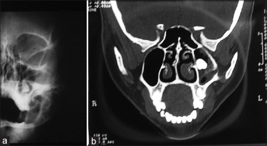
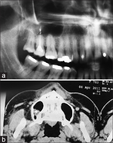
Introduction: Foreign bodies (FB) of the paranasal sinuses are an uncommon clinical entities with the maxillary sinuses being those most frequently affected. According to the literature, 60% of paranasal sinus FB are of iatrogenic origin, while 25% are of traumatic origin. This article aims to present an iatrogenic origin series of cases of FB displaced or projecting into the maxillary sinus.
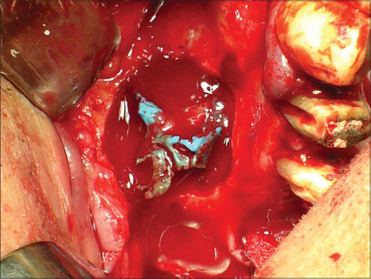
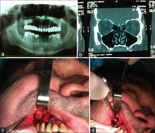
Materials and methods: In this retrospective study, the presence of the foreign body was revealed with radiologic methods and confirmed during the operation with macroscopic or later with histopathologic examination. All cases were treated with osteoplasty with vascularised pedicled bone flap or through minimally invasive intraoral procedure.
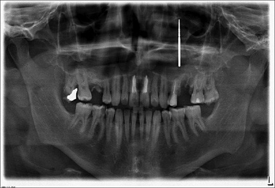
Results: A total of 27 patients were included in our study, 14 men and 13 women. The age range was 18-65 years with mean age of 46.14 (standard deviation = 10.35) years. Foreign body was displaced fragments of teeth in 11 patients (40.27%), complete teeth in four patients (14.81%), dental implants in five patients (18.51%), dental impression material in 2 cases (7,40%), gutta percha cone in two patients (7.40%), endodontic sealer associated with aspergillosis in two patients (7.40%), and dental burr in one patient (3.7%). The time between dental foreign body displacement and the surgical intervention for its removal was critical for the occurrence of sinusitis. All operated patients remained asymptomatic during a follow-up of at least 1 year.
Discussion: Prompt intervention for removal of FB eliminates the risk for chronic inflammation of the affected maxillary sinus and reduces the odds for sequelae.
Keywords: Dental implants; foreign bodies; maxillary sinus; sinusitis; tooth roots.
Copyright: © 2022 Annals of Maxillofacial Surgery.
Conflict of interest statement
There are no conflicts of interest.
Figures

Similar articles
-
[Radiologic picture of maxillary sinus aspergilloma].Otolaryngol Pol. 2010 Jun;64(7):36-9. doi: 10.1016/S0030-6657(10)70007-X. Otolaryngol Pol. 2010. PMID: 21171309 Polish.
-
An easy access to retrieve dental implants displaced into the maxillary sinus: the bony window technique.Clin Oral Implants Res. 2014 Dec;25(12):1344-51. doi: 10.1111/clr.12276. Epub 2013 Sep 30. Clin Oral Implants Res. 2014. PMID: 24112544
-
Foreign Bodies in Sinonasal Tissues: A Potential Pitfall yet a Hint to Oroantral Fistulas.Int J Surg Pathol. 2023 May;31(3):294-300. doi: 10.1177/10668969221101863. Epub 2022 May 29. Int J Surg Pathol. 2023. PMID: 35635198
-
Maxillary sinusitis caused by retained dental impression material: An unusual case report and literature review.Niger J Clin Pract. 2022 Apr;25(4):379-385. doi: 10.4103/njcp.njcp_1662_21. Niger J Clin Pract. 2022. PMID: 35439893 Review.
-
A large-scale study of treatment methods for foreign bodies in the maxillary sinus.J Oral Sci. 2018;60(3):321-328. doi: 10.2334/josnusd.18-0109. J Oral Sci. 2018. PMID: 30249933 Review.
Cited by
-
The Use of CBCT in Evaluating the Health and Pathology of the Maxillary Sinus.Diagnostics (Basel). 2022 Nov 16;12(11):2819. doi: 10.3390/diagnostics12112819. Diagnostics (Basel). 2022. PMID: 36428879 Free PMC article. Review.
References
-
- Petrov P, Daskalov H, Dzhabalyan K. Unusual case of foreign body in the maxillary sinus. Scripta Scientifica Medicinae Dentalis. 2017;3:56–8.
-
- Froum SJ, Elghannam M, Lee D, Cho SC. Removal of a dental implant displaced into the maxillary sinus after final restoration. Compend Contin Educ Dent. 2019;40:530–5. - PubMed
-
- Karpishchenko SA, Vereshchagina OE, Bolozneva EV, Karpishchenko ES. Methods of maxillary sinus foreign bodies removal. Vestn Otorinolaringol. 2020;85:78–82. - PubMed
-
- Manea C, Sarafoleanu C. Iatrogenic foreign bodies in the maxillary synus:Between malpraxis and medico-legal consequences. Rom J Legal Med. 2015;23:14.
LinkOut - more resources
Full Text Sources