

The Case of Disappearing Tibia in Rheumatoid Knee Tenosynovitis
- PMID: 36199712
- PMCID: PMC9499143
- DOI: 10.13107/jocr.2022.v12.i02.2644
The Case of Disappearing Tibia in Rheumatoid Knee Tenosynovitis
Abstract
Introduction: Knee pain and osteoarthritis are frequent patient complaints, with a rapidly increasing prevalence. By comparison, the prevalence of rheumatoid arthritis (RA) is significantly lower at around 1%. Inflammatory arthropathies, like RA, are difficult to differentiate from infection, crystal arthropathies, or malignancy. In addition, radiography and roentgenograms are often inconclusive or non-specific, making it much more difficult to evaluate, diagnose, and manage this condition. The current case is unique due to its location in the knee joint, rather than more common presentations in the upper extremities, and use of MRI imaging for diagnosis of RA with tenosynovitis.
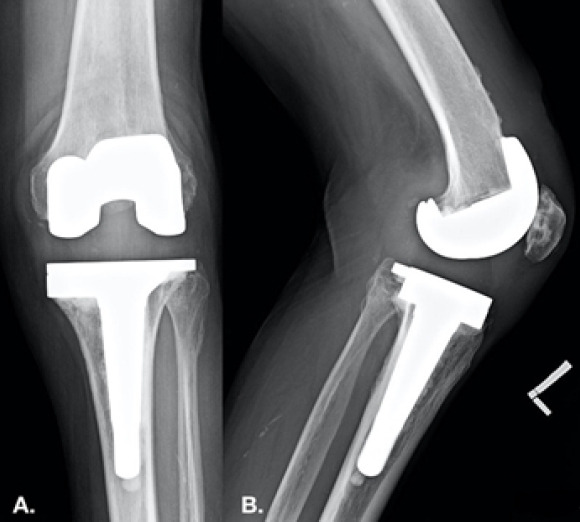
Case report: In a Caucasian 70-year-old female with sudden debilitating knee pain and a large atraumatic defect over tibial plateau, MRI showed a large fluid collection within the left gracilis muscle. Gram stain and culture of the aspirate remained negative. The only significant history involved a possible diagnosis of RA.
Conclusion: While rheumatoid tenosynovitis is common in the upper extremities, lower extremity features have not been well reported before. We diagnosed the patient with progressive RA and rheumatoid tenosynovitis. This unique presentation and rare usage of MRI imaging may be contributing to an underreporting of this diagnosis in the lower extremities.
Keywords: Rheumatoid arthritis; adult reconstruction; knee; total joint arthroplasty.
Copyright: © Indian Orthopaedic Research Group.
Conflict of interest statement
Conflict of Interest: Nil
Figures


References
-
- Jackson JL, O'Malley PG, Kroenke K. Evaluation of acute knee pain in primary care. Ann Intern Med. 2003;139:575–88. - PubMed
-
- Antoci V, Jr, Patel SP, Weaver MJ, Kwon JY. Relevance of adjacent joint imaging in the evaluation of ankle fractures. Injury. 2016;47:2366–9. - PubMed
-
- Alamanos Y, Drosos A. Epidemiology of adult rheumatoid arthritis. Autoimmun Rev. 2005;4:130–6. - PubMed
Publication types
LinkOut - more resources
Full Text Sources