

Fibrocartilaginous Dysplasia - A Report of Five Cases with Review of Literature
- PMID: 36199723
- PMCID: PMC9499150
- DOI: 10.13107/jocr.2022.v12.i02.2668
Fibrocartilaginous Dysplasia - A Report of Five Cases with Review of Literature
Abstract
Introduction: Fibrocartilaginous dysplasia (FCD) is a variant of fibrous dysplasia (FD) with extensive cartilaginous differentiation. This has been reported in both monostotic and polyostotic types of FD, the proximal femur being the most common site involved.
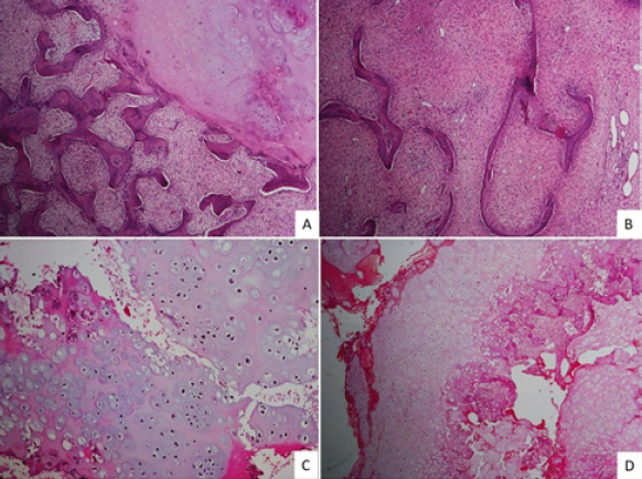
Case report: We report five cases of fibrocartilaginous dysplasia with varying degrees of cartilaginous differentiation. The age of the patients ranged from 7 to 30 years, and there was a female predominance (M:F ratio of 1:4). The proximal femur was the site of involvement in all the cases. Imaging showed well demarcated radiolucent lesions with stippled calcifications. Histologically, cartilaginous areas were noted juxtaposed to typical areas of fibrous dysplasia. Four of the patients were treated with curettage and one with a marginal resection. None of the five cases had recurrences at the past follow-up.
Conclusion: FCD is a rare variant of fibrous dysplasia which needs to diagnosed and treated early, as there is a high risk of pathological fracture.
Keywords: Fibrous dysplasia; cartilaginous tumors; chondrosarcoma; enchondroma; fibrocartilaginous.
Copyright: © Indian Orthopaedic Research Group.
Conflict of interest statement
Conflict of Interest: Nil
Figures

References
-
- Antonescu C. Soft Tissue and Bone Tumours. Lyon: International Agency for Research on Cancer; 2020.
-
- Kyriakos M, McDonald DJ, Sundaram M. Fibrous dysplasia with cartilaginous differentiation (“fibrocartilaginous dysplasia”):A review, with an illustrative case followed for 18 years. Skeletal Radiol. 2004;33:51–62. - PubMed
-
- Sanerkin NG, Watt I. Enchondromata with annular calcification in association with fibrous dysplasia. Br J Radiol. 1981;54:1027–33. - PubMed
-
- Pelzmann KS, Nagel DZ, Salyer WR. Case report 114. Skeletal Radiol. 1980;5:116–8. - PubMed
Publication types
LinkOut - more resources
Full Text Sources
Miscellaneous