

Histological Spectrum of Post Covid Debridement Tissues: Salient Histomorphological Features With Respect to Identification Fungal Elements
- PMID: 36199802
- PMCID: PMC9527210
- DOI: 10.1177/2632010X221126987
Histological Spectrum of Post Covid Debridement Tissues: Salient Histomorphological Features With Respect to Identification Fungal Elements
Abstract
Background: Secondary bacterial and fungal infections in COVID patients have been documented during current pandemic. The present study provides detailed account of histomorphology of debridement tissue received for suspected fungal infections. The primary objective was to determine the morphological characteristics that must be recognized for the identification of fungal hyphae.
Methods: The detailed histological examination of debridement tissue was performed. Demographic and clinical findings with treatment provided was recorded. Presence or absence of necrosis and lecocytoclasis was noted.
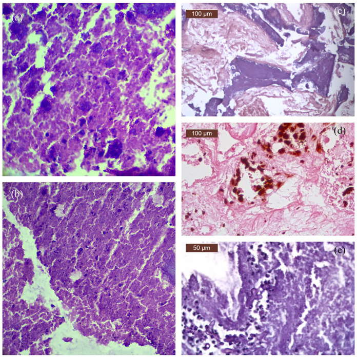
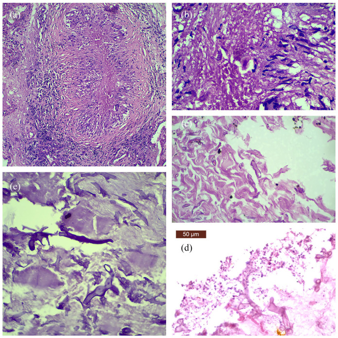
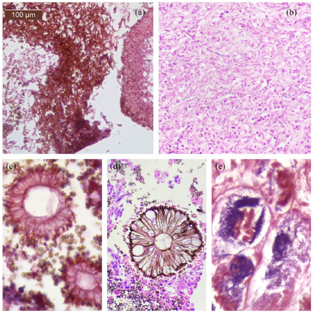
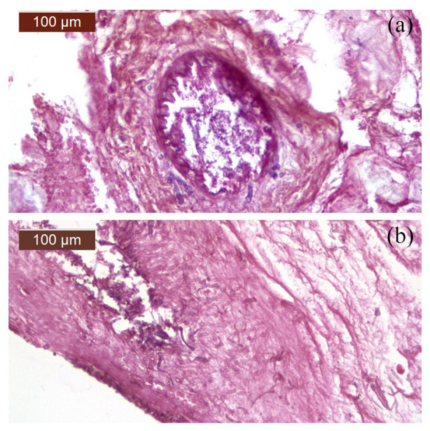
Results: A total of 110 cases of debrided tissues were included in the study. Eosinophilic granular necrosis with lecocytoclasis was observed in 103cases; fungal elements were identified in 89.3% (92/103) of these. Eleven cases where necrosis was observed, strong suspicion of fungus was reported, 6 of them displayed fungus on KOH preparation, 3 on repeat biopsy. However, in 2 of these cases, neither KOH nor repeat biopsies identified the fungus. Mucor with aspergillus was observed in 7 cases and actinomyces in 3. In all these 10 cases dense fungal colonies were evident. In 7 cases careful observation revealed fruiting bodies of aspergillus. Cotton ball appearance of actinomyces was evident. Mucor infection in current disease was so rampant that aseptate ribbon like branching mucor hyphae were evident on H&E sections. Diabetes was significantly associated with fungal infection (97.2%; 70/72; P < .005). 90% [19/21] of the patients who were on room air and diagnosed with fungal infection were diabetic.
Conclusions: Eosinophilic granular necrosis with the presence of neutrophilic debris in a case of suspected fungal disease suggests the presence of fungal elements. This warrants processing of the entire tissue deposited for examination, careful observation, application of fungal stains, and repeat biopsy if clinical suspicion is strong. Moreover, uncontrolled diabetes is more frequently associated with secondary fungal infection in COVID patients as compared to oxygen therapy.
Keywords: Covid-19; Mucor mycosis; debridement tissue; fungal infection; histology.
© The Author(s) 2022.
Conflict of interest statement
Declaration of conflicting interests: The author(s) declared no potential conflicts of interest with respect to the research, authorship, and/or publication of this article.
Figures
References
LinkOut - more resources
Full Text Sources
