. 2022 Sep 19:10:977779.
doi: 10.3389/fcell.2022.977779. eCollection 2022.

The Piwil1 N domain is required for germ cell survival in Atlantic salmon

Affiliations

The Piwil1 N domain is required for germ cell survival in Atlantic salmon

Almeida F L et al. Front Cell Dev Biol. .

Abstract

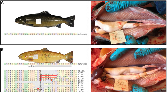
Genetic introgression of farmed salmon into wild populations can damage the genetic integrity of wild stocks and is therefore considered as an environmental threat. One possible solution is to induce sterility in farmed salmon. We have searched for proteins potentially essential for germline survival in Atlantic salmon. One of these is the argonaute protein Piwil1, known to be required for germ cell survival. To examine Piwil1 function in salmon, we induced indels in the N domain by CRISPR-Cas9. The encoded domain is present in all vertebrate Piwi proteins and has been linked to Tdrd1 protein interaction and PAZ lobe structure. The F0 founder generation of piwil1 crispant males and females displayed a mosaic pattern of piwil1 mutations, exhibiting highly mutated alleles (53%-97%) in their fin gDNA samples. In general, piwil1 crispants carried germ cells, went through puberty and became fertile, although a transient and partial germ cell loss and delays during the spermatogenic process were observed in many male crispants, suggesting that Piwil1 functions during salmon spermatogenesis. By crossing highly mutated F0 founders, we produced F1 fish with a mixture of: loss-of-function alleles (-); functional in frame mutated alleles ( + ) and wt alleles (+). In F1, all piwil1 -/- fish lacked germ cells, while piwil1 +/+ siblings showed normal ovaries and testes. Yet, most juvenile F1 piwil1 +/-males and females displayed an intermediate phenotype with a higher somatic/germ cell ratio without an increase in germ cell apoptosis, suggestive of a gene dose effect on the number of germ cells and/or insufficient replacement of lost germ cells in heterozygous fish. Interestingly, the two longest in-frame indels in the N domain also ensured germ cell loss. Hence, the loss of 4-6 aa in this region Phe130-Ser136 may result in crucial changes of the protein structure, potentially affecting piRNA binding of the PAZ lobe, and/or affecting the binding of Piwil1 interacting proteins such as Tdrd protein, with critical consequences for the survival of primordial germ cells. In conclusion, we show that loss of piwil1 leads to loss of germ cells in salmon and that part of the N domain of Piwil1 is crucial for its function.

Keywords: CRISPR/Cas9; argonaute protein (AGO); fish sterility; germline; spermatogenesis.


Conflict of interest statement

The authors declare that the research was conducted in the absence of any commercial or financial relationships that could be construed as a potential conflict of interest.

Figures

FIGURE 1
FIGURE 1
Gonadosomatic index (GSI; %) and testis histology of wild type (wt) and piwil1 F0 crispant Atlantic salmon: (A) 24 January 2017: males of both genotypes were immature, as indicated by spermatogenic tubules containing only Sertoli cells and type A spermatogonia. The testes of 3 out of 5 crispants showed germ cell-free areas containing only Sertoli cells (see area to the right of the stippled line). The apparent frequency of cells in mitosis (black arrows) was lower in crispant testes that also showed several areas in which Sertoli cell groups were not in contact with germ cells (black delineated areas in A′); (B) 21 February 2017: puberty had started in both genotypes, as indicated by the presence of type B spermatogonia (SgB); apoptotic cells (white delineated areas) were found frequently in crispant testes but rarely in wt testes; (C) 9 May 2017: rapid testicular growth phase reflecting full spermatogenic activity, maturing crispants showed a lower GSI, apparently less spermatozoa (Sz) and apoptotic cells (white delineated areas) than wt males. (D) 26 September 2017: the large majority of both wt and crispant males were spermiating 599 and the lumina of the spermatogenic tubules were filled with spermatozoa (Sz), while the germinal 600 epithelium consisted of a single layer of Sertoli cells (white arrows) and scattered single, apparently 601 quiescent type A spermatogonia (black circles). Black arrows: mitosis; white-delineated areas: 602 apoptotic germ cells; white arrows: Sertoli cells. Immat—immature, mat—maturing. Sc—603 spermatocyte, SgB—type B spermatogonia, St—spermatid, Sz—spermatozoa.
FIGURE 2
FIGURE 2
External appearance and macroscopic testis anatomy of 1 year old wt [(A); animal #79) and piwil1 F0 crispant Atlantic salmon [(B); animal #80) males. The table in B shows an alignment of the most common (up to 2%) piwil1 mutations found in #80. Red rectangles show inserted bases and bold letters indicate nucleotide substitutions.
FIGURE 3
FIGURE 3
Plasma levels of 11-ketotestosterone (11KT; ng/ml) of 1 year old male Atlantic salmon (control - wt and piwil1 F0 crispant). Ma-maturing, im–immature.
FIGURE 4
FIGURE 4
External appearance and macroscopic view of ovulated eggs of 2 years old wt [(A); animal #127) and piwil1 F0 crispant Atlantic salmon [(B); animal #132) females. The table in B shows an alignment of the most common (up to 2%) piwil1 mutations found in #132. Bold letters indicate nucleotide substitutions.
FIGURE 5
FIGURE 5
Alignment and frequencies of indels in piwil1 F0 and F1 Atlantic salmon. (A) Alignment of the sequences identified in the piwil1 crispant F0 salmon used as breeders to produce the F1 generation. In bold, indels that lead to loss of function in homozygosis. Red arrows point to wt piwil1 copy (present only in the males). Indels with frequency lower than 2% were not included in the figure. Red rectangles show insertions and bold letters indicate base substitutions. (B) Frequency of indels found in piwil1 +/− and piwil1 −/− F1 salmon. F: female; M: male.
FIGURE 6
FIGURE 6
Gonads of wild type (wt) and piwil1 F1 KO (−/−) Atlantic salmon. (A) Testis of a wt male. (B) Testis of a piwil1 KO (del20/del5) male (#298). (C) Ovary of a wt female. (D) Ovary of a piwil1 KO (del8/del12) female (#225). White arrows point to Sertoli cells; red arrows show type A spermatogonia; white asterisks mark dense interstitium and red asterisks mark interstitium rich in fibrocytes and extracellular matrix; white line marks groups of follicle cells. Oc: previtellogenic oocyte.
FIGURE 7
FIGURE 7
Phenotype and genotype of all piwil1 F1 Atlantic salmon sampled on six occasions. Fish displayed germ cell free gonads (GCF), intermediate phenotype or normal gonads. Data from September 2020 combines two samplings in the same month (9th and 16th).
FIGURE 8
FIGURE 8
Gonads of piwil1 +/+ (wt) and piwil1 +/− F1 Atlantic salmon. (A) Testis of a wild type (wt) male. (B)Testis of a piwil1 +/− (wt/del8) male presenting a lower GC/somatic cells ratio. (C) Ovary of a wt female. (D) Ovary of a piwil1 +/− (del3/del8) female with excess of early germ cells. Black arrows point to Sertoli cell nuclei; red arrows show nuclei of type A spermatogonia. Continuous line marks groups of oogonia and stripled line marks cystic oocytes. Oo: previtellogenic oocyte.
FIGURE 9
FIGURE 9
Fold relative expression of genes expressed in the gonads and gonadosomatic indices of wild type (wt), piwil1 +/− and piwil1 +/− F1 Atlantic salmon. (A) gsdf and vasa in testes. (B) amh and vasa in testes. (C) cyp19a1a and vasa in ovaries. (D) Gonadosomatic index (GSI) in wt, piwil1 +/− and piwil1 −/− F1 Atlantic salmon males and females.
FIGURE 10
FIGURE 10
Homology modelling of the inframe lof alleles del12 and del18. Top row displays the wildtype modelled salmon Piwi1 with deleted amino acids shown in purple. The red coloration is reflecting the positional root-mean-square deviation (RMSD) between two sets of atoms in the mutant versus wildtype. The bottom row displays the modelled structure of the variants with the red coloration indicating the RMSD value.

References

    1. Ayllon F., Solberg M. F., Glover K. A., Mohammadi F., Kjaerner-Semb E., Fjelldal P. G., et al. (2019). The influence of vgll3 genotypes on sea age at maturity is altered in farmed mowi strain Atlantic salmon. BMC Genet. 20, 44. 10.1186/s12863-019-0745-9 - DOI - PMC - PubMed
    1. Carmell M. A., Girard A., van de Kant H. J., Bourc'his D., Bestor T. H., de Rooij D. G., et al. (2007). MIWI2 is essential for spermatogenesis and repression of transposons in the mouse male germline. Dev. Cell 12 (4), 503–514. 10.1016/j.devcel.2007.03.001 - DOI - PubMed
    1. Clement K., Rees H., Canver M. C., Gehrke J. M., Farouni R., Hsu J. Y., et al. (2019). CRISPResso2 provides accurate and rapid genome editing sequence analysis. Nat. Biotechnol. 37, 224–226. 10.1038/s41587-019-0032-3 - DOI - PMC - PubMed
    1. Conant D., Hsiau T., Rossi N., Oki J., Maures T., Waite K., et al. (2022). Inference of CRISPR edits from sanger trace data. CRISPR J. 5 (1), 123–130. 10.1089/crispr.2021.0113 - DOI - PubMed
    1. Cox D. N., Chao A., Lin H. (2000). Piwi encodes a nucleoplasmic factor whose activity modulates the number and division rate of germline stem cells. Development 127 (3), 503–514. 10.1242/dev.127.3.503 - DOI - PubMed

LinkOut - more resources