

Case Reports
doi: 10.1155/2022/2104120.
eCollection 2022.
A Rare Case of Extensive Eggshell Intestinal Wall Peritoneal Calcification in a Long-Term Continuous Peritoneal Dialysis Patient
Affiliations
- PMID: 36200067
- PMCID: PMC9529488
- DOI: 10.1155/2022/2104120
Item in Clipboard
Case Reports
A Rare Case of Extensive Eggshell Intestinal Wall Peritoneal Calcification in a Long-Term Continuous Peritoneal Dialysis Patient
Case Rep Nephrol.
.
Abstract
Encapsulating peritoneal sclerosis (EPS) is a rare but rather serious complication of long-term peritoneal dialysis. The etiology of EPS is multifactorial, with long-term peritoneal dialysis, multiple peritonitis episodes, and uncontrolled hyperparathyroidism considered to be major risk factors for this often life-threatening condition. We report a case of a 55-year-old female patient with Down syndrome and end-stage renal disease (ESRD) on long-term peritoneal dialysis (PD) with extensive intestinal peritoneal calcifications and a rather uncomplicated long follow-up.
Copyright © 2022 Erasmia Sampani et al.
Conflict of interest statement
The authors declare that they have no conflicts of interest.
Figures
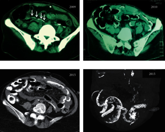
Abdominal CT scans of the patient in 2009 (a), showing mild linear peritoneal calcifications (arrows), which became full-blown calcifications of the visceral small bowel peritoneum a year later in 2010 (b) that remained or minimally increased over the next five years (2015 (c)). (d) Processed sagittal plane image of the 2015 abdominal scan showing the extent of bowel wall calcifications.
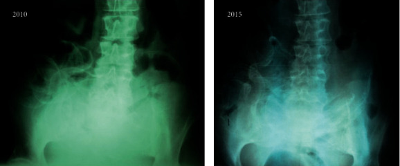
Plain abdominal films of the patient, without any contrast media, showing extensive calcifications of the bowel wall in 2010 (a) and five years later, in 2015 (b).
References
-
- Nomoto Y., Kawaguchi Y., Kubo H., Hirano H., Sakai S., Kurokawa K. Sclerosing encapsulating peritonitis in patients undergoing continuous ambulatory peritoneal dialysis: a report of the Japanese sclerosing encapsulating peritonitis study group. American Journal of Kidney Diseases . 1996;28(3):420–427. doi: 10.1016/s0272-6386(96)90501-6. - DOI - PubMed
Publication types
LinkOut - more resources
Full Text Sources
