Review
. 2022 Sep 19:9:1008175.
doi: 10.3389/fmolb.2022.1008175. eCollection 2022.

Non-lysine ubiquitylation: Doing things differently

Affiliations
Review

Non-lysine ubiquitylation: Doing things differently

Ian R Kelsall. Front Mol Biosci. .

Abstract

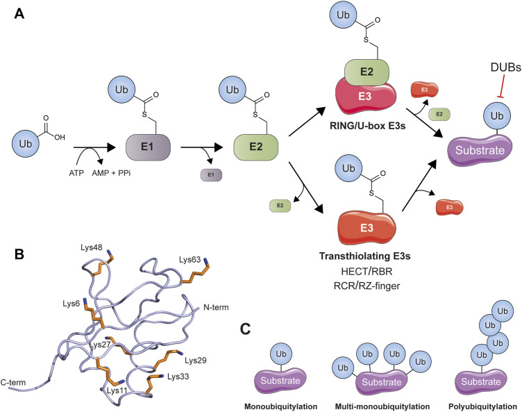
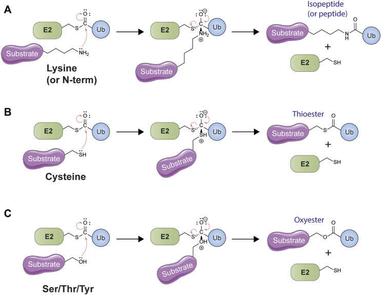
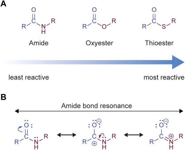
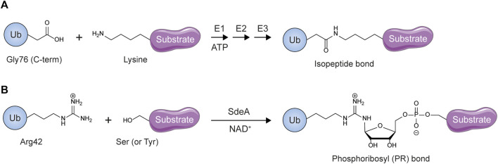
The post-translational modification of proteins with ubiquitin plays a central role in nearly all aspects of eukaryotic biology. Historically, studies have focused on the conjugation of ubiquitin to lysine residues in substrates, but it is now clear that ubiquitylation can also occur on cysteine, serine, and threonine residues, as well as on the N-terminal amino group of proteins. Paradigm-shifting reports of non-proteinaceous substrates have further extended the reach of ubiquitylation beyond the proteome to include intracellular lipids and sugars. Additionally, results from bacteria have revealed novel ways to ubiquitylate (and deubiquitylate) substrates without the need for any of the enzymatic components of the canonical ubiquitylation cascade. Focusing mainly upon recent findings, this review aims to outline the current understanding of non-lysine ubiquitylation and speculate upon the molecular mechanisms and physiological importance of this non-canonical modification.

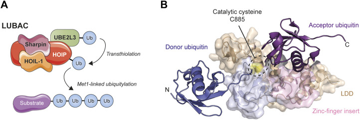
Keywords: ERAD; LUBAC; Rnf213; non-canonical ubiquitylation; non-lysine ubiquitylation; oxyester bond; thioester bond; ubiquitin.


Conflict of interest statement

The author declares that the research was conducted in the absence of any commercial or financial relationships that could be construed as a potential conflict of interest.

Figures

FIGURE 1
FIGURE 1
Components of the conventional ubiquitylation cascade. (A) A multicomponent enzyme cascade is used to transfer ubiquitin (Ub) to its substrates. RING and U-box ligases facilitate the transfer of ubiquitin from E2 directly to substrate lysine whereas the transthiolating classes of ligase (HECT/RBR/RCR/RZ-finger) first covalently bind ubiquitin at their own active site cysteine before transferring it to substrate. Ubiquitin can be removed by specialized deubiquitylating enzymes (DUBs). (B) The structure of ubiquitin (PDB: 1UBQ) (Vijay-Kumar et al., 1987) is shown as a smoothed backbone trace. The position of ubiquitin’s seven lysine residues is indicated. (C) Different ubiquitylation topologies are possible: monoubiquitylation, multi-monoubiquitylation, and polyubiquitylation (in which ubiquitin can ubiquitylate itself to form amide-linked chains) all occur.
FIGURE 2
FIGURE 2
Canonical and non-canonical ubiquitylation both represent examples of nucleophilic acyl transfer. (A) Conventional ubiquitylation involves the attack of the electrophilic thioester carbonyl of an E2-ubiquitin (Ub) conjugate by the lone pair electrons of the substrate lysine amine group. This leads to the formation and then collapse of a tetrahedral intermediate, expelling the E2 leaving group and resulting in lysine ubiquitylation. The same mechanism may also be employed to ubiquitylate the free amine group of the N-terminus of the polypeptide backbone. (B) Like nitrogen, the cysteine thiol also possesses a lone pair of electrons and can act as a nucleophile in an acyl transfer reaction. This same mechanism is employed by E2s themselves when they accept ubiquitin from the E1 enzyme, as well as by the transthiolating E3 ligases. This reaction leads to the formation of a thioester. (C) The amino acids serine, threonine, and tyrosine possess a nucleophilic hydroxyl group which can be ubiquitylated by means of a nucleophilic acyl transfer mechanism. The resulting linkage between ubiquitin and the substrate is an oxyester.
FIGURE 3
FIGURE 3
Met1-linked ubiquitin is synthesized by the Linear Ubiquitin Chain Assembly Complex (LUBAC). (A) LUBAC is a trimeric complex composed of HOIP, HOIL-1, and Sharpin. HOIP binds its cognate E2 (very likely UBE2L3 (Lewis et al., 2015)) and by means of a transthiolation mechanism that sees ubiquitin transferred to Cys885 in the catalytic RBR domain of the ligase, builds Met1-linked (‘linear’) ubiquitin chains in which peptide bonds between the C- and N-termini of ubiquitin create a single continuous polypeptide chain of repeating ubiquitin units. (B) Structure of the minimal catalytic core of HOIP in complex with acceptor (purple) and donor (dark blue) ubiquitin (PDB: 4LJO) (Stieglitz et al., 2013). The unique LDD (wheat colour) and zinc-finger (pink) domains of HOIP help ensure that the C-terminus of the donor ubiquitin and N-terminus of the acceptor ubiquitin align for Met1 linkage formation. Note the proximity of the catalytic cysteine (coloured yellow) to the N-terminus of the acceptor ubiquitin, explaining the specificity for linear chains.
FIGURE 4
FIGURE 4
Novel sequence features of UBE2W and UBE2J2 that may determine non-canonical specificity. (A) A sequence alignment of the residues in selected human and yeast E2s close to the active site cysteine (highlighted in red) reveals that canonical lysine-directed E2s contain a conserved upstream arginine residue (normally part of an HPN motif) and a downstream Asp/Ser residue (both highlighted in yellow). E2s known to ubiquitylate hydroxylated residues are written in red and notably differ in the amino acids present at these sites. Divergent catalytically-important amino acids in UBE2W are highlighted in green, including the basic cluster that replaces the conserved D/S residue. The equivalent residues in UBE2J2 and its yeast homologue UBC6 are highlighted in blue. The names of E2s involved in transthiolation of cysteine residues are written in orange. Sequence alignments were performed using the Clustal Omega server (Sievers and Higgins 2021) and manually edited where necessary to match known structural information (Gundogdu and Walden 2019). (B) Phylogenetic tree depicting relationships between the selected E2s shown in (A). The non-canonical ERAD-related E2 conjugating enzyme UBE2J2 and its yeast homologue UBC6 are highlighted in blue, UBE2W is highlighted in green. Analysis was performed by aligning the ubiquitin-conjugating UBC fold of selected E2s in Clustal Omega and is displayed using iTOL (Letunic and Bork 2021).
FIGURE 5
FIGURE 5
The relative reactivity of ubiquitin conjugating bonds. (A) The amide group is the least reactive, and therefore the most stable, of the acyl groups formed during ubiquitylation. The thioester is the most reactive, and therefore the least stable, whereas the oxyester is somewhere in between. (B) The stability of the amide bond is due to stabilization of the partial positive charge on the carbonyl carbon by electron donation from non-bonding electrons on the adjacent nitrogen, thus decreasing electrophilicity. In contrast, thioesters exhibit much weaker resonance stabilization due to poor overlap between the electron orbitals in sulfur and carbon.
FIGURE 6
FIGURE 6
Membrane-spanning E3 ligase complexes facilitate non-lysine ubiquitylation during PEX5 recycling and ER-associated protein degradation. (A) Cargo proteins destined for peroxisomal import are recognized in the cytosol by PEX5. PEX5 and cargo then translocate from the cytosol into the lumen of the peroxisome. The unstructured N-terminus of PEX5 inserts into the pore of an E3 ligase complex containing three RING-type E3s and is monoubiquitylated on a cysteine residue, triggering the extraction of PEX5 from the peroxisome by a hexameric ATPase. This extraction is accompanied by PEX5 unfolding and cargo release. Once in the cytosol PEX5 refolds and is deubiquitylated, allowing a new round of cargo import to begin. (B) Misfolded or unfolded glycoproteins in the ER lumen are retrotranslocated into the cytosol by an E3 ligase complex and ubiquitylated by this complex upon emergence on the cytosolic side. The membrane-bound ubiquitin conjugating enzyme UBE2J2 acts with the ligase complex to modify a range of amino acid residues (Lys/Cys/Ser/Thr) in the misfolded target protein, triggering its proteasomal degradation.
FIGURE 7
FIGURE 7
HOIL-1 catalyzes serine and threonine ubiquitylation. (A) Structure of ubiquitin (PDB: 1UBQ) (Vijay-Kumar et al., 1987) highlighting the eleven hydroxylated amino acids (shown in red). HOIL-1 can form ubiquitin dimers in vitro, with oxyester linkages at Thr12, Ser20, Thr22, and Thr55 all reported. Note that the last three of these residues cluster at a single region on the surface of ubiquitin. (B) Alignment of the human RBR domain sequences in the catalytic RING2 domain. Conserved zinc coordinating residues that form the RING2 fold are highlighted yellow, the active site cysteine is highlighted in red, the catalytic histidine residue present in most RBRs is highlighted in gray; in HOIL-1 this is a tryptophan and is highlighted in blue. Unique inserts exist in HOIL-1, HOIP, and RNF216, and putative or confirmed zinc coordinating residues are highlighted purple. The identity of the last three zinc binding residues in the HOIL-1 RING domain is currently unknown and consequently these residues are not aligned.
FIGURE 8
FIGURE 8
Non-proteinaceous substrates of ubiquitylation. (A) The Lipid A component of bacterial lipopolysaccharide represents the minimal substrate for RNF213-mediated ubiquitylation of LPS. The precise site of attachment is not yet known. (B) Glycogen is a highly branched polymer of glucose. Most glucose units are linked together by α(1,4)-glycosidic bonds with branches formed by the introduction of glycosidic bonds at the C6 hydroxy position every 8–12 glucose units. This C6 hydroxy group is also the target of HOIL-1-catalysed ubiquitylation of glycogen and related sugars. (C) The Deltex family E3 ligases can catalyze the conjugation of ubiquitin to ADP-ribose by means of an oxyester linkage between the 1′-hydroxy group of ribose and ubiquitin’s C-terminus. This reaction requires all the components of the ubiquitylation cascade and involves no recognized ADP-ribosyltransferase.
FIGURE 9
FIGURE 9
Unconventional ubiquitylation by Legionella pneumophilia. (A) Conventional eukaryotic ubiquitylation modifies substrate lysines. (B) PR-ubiquitylation is catalyzed by SidE effectors such as SdeA and targets substrate serines and tyrosines.

Similar articles

Cited by

References

    1. Adalbert R., Coleman M. P. (2013). Review: Axon pathology in age-related neurodegenerative disorders. Neuropathol. Appl. Neurobiol. 39, 90–108. 10.1111/j.1365-2990.2012.01308.x - DOI - PubMed
    1. Ahel J., Fletcher A., Grabarczyk D. B., Roitinger E., Deszcz L., Lehner A., et al. (2021). E3 ubiquitin ligase RNF213 employs a non-canonical zinc finger active site and is allosterically regulated by ATP. bioRxiv 2021, 443411. 10.1101/2021.05.10.443411 - DOI
    1. Ahel J., Lehner A., Vogel A., Schleiffer A., Meinhart A., Haselbach D., et al. (2020). Moyamoya disease factor RNF213 is a giant E3 ligase with a dynein-like core and a distinct ubiquitin-transfer mechanism. Elife 9, e56185. 10.7554/eLife.56185 - DOI - PMC - PubMed
    1. Ahmed S. F., Buetow L., Gabrielsen M., Lilla S., Chatrin C., Sibbet G. J., et al. (2020). DELTEX2 C-terminal domain recognizes and recruits ADP-ribosylated proteins for ubiquitination. Sci. Adv. 6, eabc0629. 10.1126/sciadv.abc0629 - DOI - PMC - PubMed
    1. Akimov V., Barrio-Hernandez I., Hansen S. V. F., Hallenborg P., Pedersen A. K., Bekker-Jensen D. B., et al. (2018a). UbiSite approach for comprehensive mapping of lysine and N-terminal ubiquitination sites. Nat. Struct. Mol. Biol. 25, 631–640. 10.1038/s41594-018-0084-y - DOI - PubMed

LinkOut - more resources