

Ultra-low Concentration of Cellulose Nanofibers (CNFs) for Enhanced Nucleation and Yield of ZnO Nanoparticles
- PMID: 36200128
- PMCID: PMC9583615
- DOI: 10.1021/acs.langmuir.2c01713
Ultra-low Concentration of Cellulose Nanofibers (CNFs) for Enhanced Nucleation and Yield of ZnO Nanoparticles
Abstract
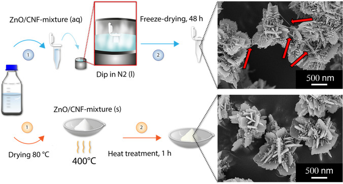
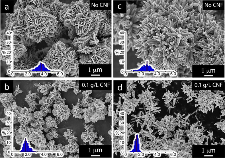
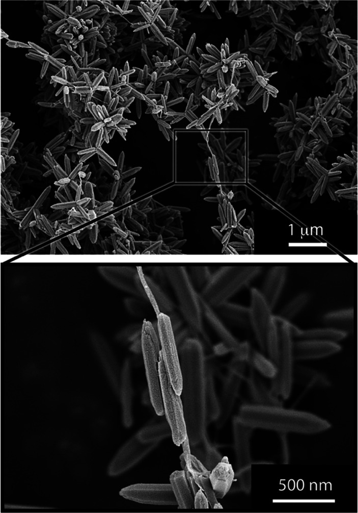
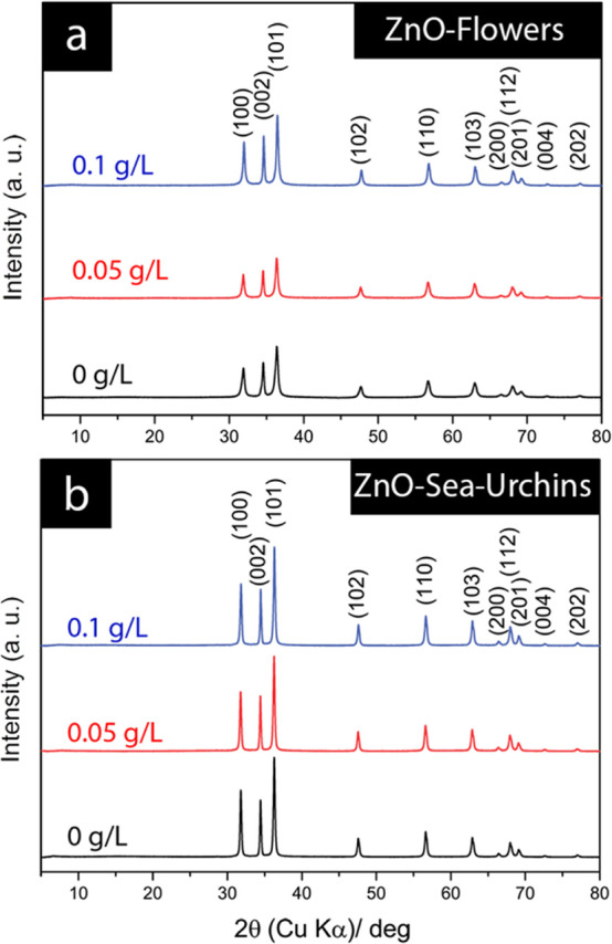
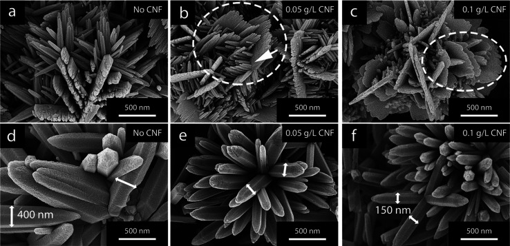
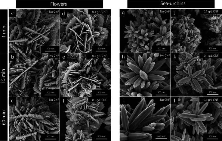
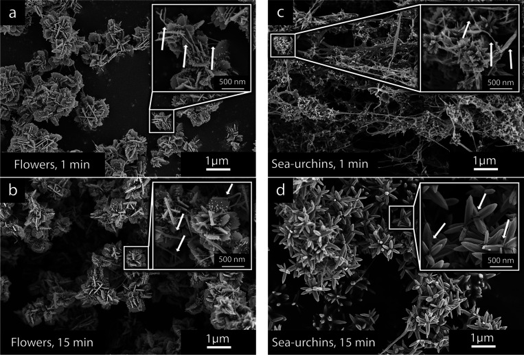
Cellulose nanofibers (CNFs) were used in aqueous synthesis protocols for zinc oxide (ZnO) to affect the formation of the ZnO particles. Different concentrations of CNFs were evaluated in two different synthesis protocols producing distinctly different ZnO morphologies (flowers and sea urchins) as either dominantly oxygen- or zinc-terminated particles. The CNF effects on the ZnO formation were investigated by implementing a heat-treatment method at 400 °C that fully removed the cellulose material without affecting the ZnO particles made in the presence of CNFs. The inorganic phase formations were monitored by extracting samples during the enforced precipitations to observe changes in the ZnO morphologies. A decrease in the size of the ZnO particles could be observed for all synthesis protocols, already occurring at small additions of CNFs. At as low as 0.1 g/L CNFs, the particle size decreased by 50% for the flower-shaped particles and 45% for the sea-urchin-shaped particles. The formation of smaller particles was accompanied by increased yield by 13 and 15% due to the CNFs' ability to enhance the nucleation, resulting in greater mass of ZnO divided among a larger number of particles. The enhanced nucleation could also be verified as useful for preventing secondary morphologies from forming, which grew on the firstly precipitated particles. The suppression of secondary growths' was due to the more rapid inorganic phase formation during the early phases of the reactions and the faster consumption of dissolved salts, leaving smaller amounts of metal salts present at later stages of the reactions. The findings show that using cellulose to guide inorganic nanoparticle growth can be predicted as an emerging field in the preparation of functional inorganic micro/nanoparticles. The observations are highly relevant in any industrial setting for the large-scale and resource-efficient production of ZnO.
Conflict of interest statement
The authors declare no competing financial interest.
Figures
Similar articles
-
Functional biocompatible nanocomposite films consisting of selenium and zinc oxide nanoparticles embedded in gelatin/cellulose nanofiber matrices.Int J Biol Macromol. 2021 Apr 1;175:87-97. doi: 10.1016/j.ijbiomac.2021.01.135. Epub 2021 Jan 22. Int J Biol Macromol. 2021. PMID: 33485892
-
An Organic/Inorganic Nanocomposite of Cellulose Nanofibers and ZnO Nanorods for Highly Sensitive, Reliable, Wireless, and Wearable Multifunctional Sensor Applications.ACS Appl Mater Interfaces. 2019 Dec 26;11(51):48239-48248. doi: 10.1021/acsami.9b17824. Epub 2019 Dec 12. ACS Appl Mater Interfaces. 2019. PMID: 31766842
-
The Fast and One-Step Growth of ZnO Nanorods on Cellulose Nanofibers for Highly Sensitive Photosensors.Nanomaterials (Basel). 2023 Sep 21;13(18):2611. doi: 10.3390/nano13182611. Nanomaterials (Basel). 2023. PMID: 37764641 Free PMC article.
-
Cellulose from sources to nanocellulose and an overview of synthesis and properties of nanocellulose/zinc oxide nanocomposite materials.Int J Biol Macromol. 2020 Jul 1;154:1050-1073. doi: 10.1016/j.ijbiomac.2020.03.163. Epub 2020 Mar 19. Int J Biol Macromol. 2020. PMID: 32201207 Review.
-
Review on Nonconventional Fibrillation Methods of Producing Cellulose Nanofibrils and Their Applications.Biomacromolecules. 2021 Oct 11;22(10):4037-4059. doi: 10.1021/acs.biomac.1c00640. Epub 2021 Sep 10. Biomacromolecules. 2021. PMID: 34506126 Review.
Cited by
-
Sprayed water-based lignin colloidal nanoparticle-cellulose nanofibril hybrid films with UV-blocking ability.Nanoscale Adv. 2024 Aug 28;6(20):5031-41. doi: 10.1039/d4na00191e. Online ahead of print. Nanoscale Adv. 2024. PMID: 39247863 Free PMC article.
References
-
- Benitez A. J.; Walther A. Cellulose nanofibril nanopapers and bioinspired nanocomposites: a review to understand the mechanical property space. J. Mater. Chem. A 2017, 5, 16003–16024. 10.1039/C7TA02006F. - DOI
-
- Zhai L.; Kim H. C.; Kim J. W.; Kang J.; Kim J. Elastic moduli of cellulose nanofibers isolated from various cellulose resources by using aqueous counter collision. Cellulose 2018, 25, 4261–4268. 10.1007/s10570-018-1836-x. - DOI
-
- Moser C.; Backlund H.; Drenth L.; Henriksson G.; Lindstrom M. E. Xyloglucan adsorption for measuring the specific surface area on various never-dried cellulose nanofibers. Nord. Pulp Pap. Res. J. 2018, 33, 186–193. 10.1515/npprj-2018-3034. - DOI
Publication types
MeSH terms
Substances
LinkOut - more resources
Full Text Sources
Miscellaneous
