. 2023 Apr 19;146(4):1373-1387.
doi: 10.1093/brain/awac364.

Loss-of-function variants in MYCBP2 cause neurobehavioural phenotypes and corpus callosum defects

Affiliations

Loss-of-function variants in MYCBP2 cause neurobehavioural phenotypes and corpus callosum defects

Lama AlAbdi et al. Brain. .

Abstract

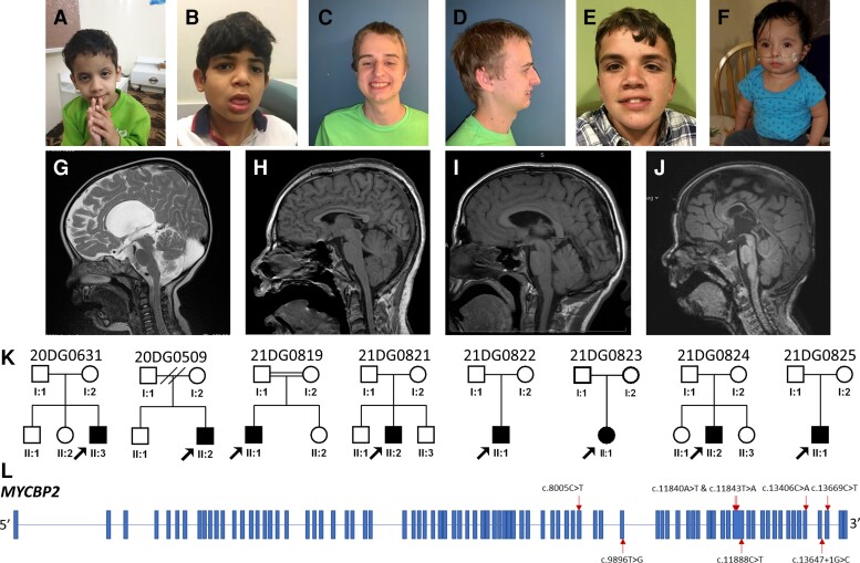
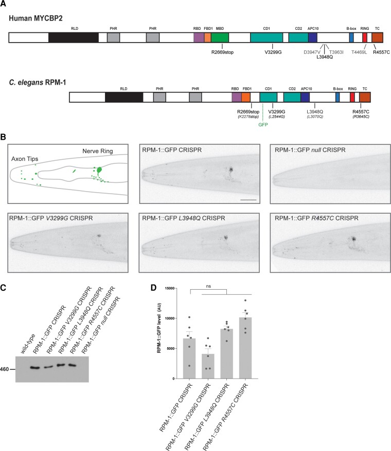
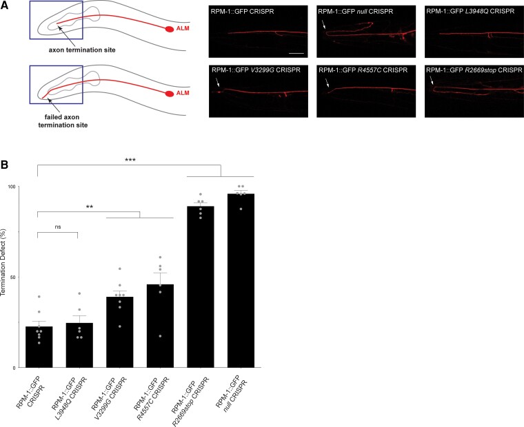
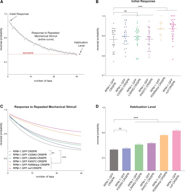
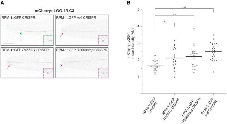
The corpus callosum is a bundle of axon fibres that connects the two hemispheres of the brain. Neurodevelopmental disorders that feature dysgenesis of the corpus callosum as a core phenotype offer a valuable window into pathology derived from abnormal axon development. Here, we describe a cohort of eight patients with a neurodevelopmental disorder characterized by a range of deficits including corpus callosum abnormalities, developmental delay, intellectual disability, epilepsy and autistic features. Each patient harboured a distinct de novo variant in MYCBP2, a gene encoding an atypical really interesting new gene (RING) ubiquitin ligase and signalling hub with evolutionarily conserved functions in axon development. We used CRISPR/Cas9 gene editing to introduce disease-associated variants into conserved residues in the Caenorhabditis elegans MYCBP2 orthologue, RPM-1, and evaluated functional outcomes in vivo. Consistent with variable phenotypes in patients with MYCBP2 variants, C. elegans carrying the corresponding human mutations in rpm-1 displayed axonal and behavioural abnormalities including altered habituation. Furthermore, abnormal axonal accumulation of the autophagy marker LGG-1/LC3 occurred in variants that affect RPM-1 ubiquitin ligase activity. Functional genetic outcomes from anatomical, cell biological and behavioural readouts indicate that MYCBP2 variants are likely to result in loss of function. Collectively, our results from multiple human patients and CRISPR gene editing with an in vivo animal model support a direct link between MYCBP2 and a human neurodevelopmental spectrum disorder that we term, MYCBP2-related developmental delay with corpus callosum defects (MDCD).

Keywords: MYCBP2; Phr1; corpus callosum; epilepsy; habituation; neurodevelopmental disorder.


Conflict of interest statement

The Department of Molecular and Human Genetics at Baylor College of Medicine receives revenue from clinical genetic testing completed at Baylor Genetics Laboratories. K.M. and A.B. are employees of GeneDx, Inc. N. Z. is a co-founder and Scientific Advisory Board Member of Coho Therapeutics Inc. and SEED Therapeutics Inc. The remaining authors declare no competing interests.

Figures

Figure 1
Figure 1
Overview of patients with MYCBP2 variants identified in this study. (AG) Clinical pictures highlighting the variable facial dysmorphisms in patients (see Supplementary Table 1 for further details): (A) 20DG0631, (B) 21DG0819, (C and D) 21DG0821, (E) 20DG0509, and (F) 21DG0825. (GK) Sagittal MRI images highlighting the range of corpus callosum defects in the study cohort: (G) 20DG0631, with agenesis of corpus callosum, (H and I) 21DG0819 and 20DG0509, respectively, with mild thinning of the corpus callosum, (J) 21DG0822 with dysgenesis of the corpus callosum. (K) Pedigrees for all the patients participating in this study. (L) Schematic of MYCBP2 with variants annotated.
Figure 2
Figure 2
CRISPR editing MDCD-related variants into rpm-1 does not impair protein localization or stability in C. elegans. (A) Schematic of human MYCBP2 and its C. elegans orthologue RPM-1. Shown are protein sequence changes caused by MYCBP2 missense and nonsense variants from MDCD patients. Also noted is the location of the GFP inserted using CRISPR/Cas9 gene engineering in C. elegans and annotated protein domains. RCC1 like domain (RLD), Pam/Highwire/RPM-1 (PHR) domain, RAE-1 binding domain (RBD), FSN-1 binding domain (FBD), conserved domain of unknown function (CD), anaphase-promoting complex subunit 10 like domain (APC10). (B) CRISPR engineering was used to fuse GFP with endogenous RPM-1. CRISPR editing then introduced conserved MDCD-related variants (V3299G, L3948Q and R4557C) or a known null mutation (C4587Y) into RPM-1::GFP CRISPR animals. Shown is a schematic of RPM-1::GFP CRISPR localization at axon tips and the nerve ring in the head of C. elegans (left). Representative confocal images are shown for all genotypes. Note localization and levels of RPM-1::GFP are normal in CRISPR edited animals carrying MDCD-related variants (V3299G, L3948Q and R4557C). (C) Immunoprecipitation of RPM-1::GFP from whole C. elegans lysates for indicated genotypes. Representative of three independent experiments. (D) Quantitation of RPM-1::GFP for indicated genotypes. Means are shown for six replicates from three independent experiments for each genotype. Error bars represent SEM. Significance determined using Student’s t-test with Bonferroni correction. ns = not significant. Scale bar = 20 μm.
Figure 3
Figure 3
MDCD-related variants affect in vivo axon development in C. elegans. (A) Top: Axon termination site (arrow) for ALM mechanosensory neuron of C. elegans. Bottom: Failed termination (arrow) observed in MDCD-related variants. Shown are representative images of ALM axons for indicated genotypes visualized using transgenic, cell-specific RFP. Axon termination defects (arrow) are observed in RPM-1::GFP null CRISPR animals and in RPM-1::GFP CRISPR animals harbouring MDCD-related variants (V3299G, R4557C and R2669stop). (B) Quantitation of axon termination defects for indicated genotypes. Means are shown from six to eight counts (20–30 animals per count) for each genotype, and error bars represent SEM. Significance determined using Student’s t-test. **P < 0.01; ***P < 0.001; ns = not significant. Scale bar = 20 μm.
Figure 4
Figure 4
C. elegans behavioural responses are altered by MDCD-related variants. (A) Multiple behavioural responses to mechanical stimulation were evaluated using computationally automated assays in C. elegans. Shown is raw control data (boxes) for RPM-1::GFP CRISPR animals and analysis points for initial response, response to repeated mechanical stimulation, and habituation level. Exponential fit curve across assays (solid line) determines habituation in two ways, response to repeated mechanical stimulation (overall curve) and habituation level (value of fit curve asymptote, dashed line). (B) Quantitation of initial response for indicated genotypes. RPM-1::GFP CRISPR animals carrying further CRISPR edits for MDCD-related missense variants have normal initial responses to mechanical stimulation. RPM-1::GFP R2669stop and null CRISPR animals show higher sensitivity to initial stimulation. (C) Quantitation showing all MDCD-related variants display abnormalities in response to repeated mechanical stimulation. See Supplementary Fig. 7 for raw data plots for all genotypes. (D) Quantitation of habituation levels for indicated genotypes. Habituation is impaired in RPM-1::GFP CRISPR animals carrying L3948Q, R4557C, and R2669stop MDCD-related variants. For C and D, averages are shown from 29–30 replicates (60–100 animals per replicate) for each genotype obtained from eight independent experiments, except for R2669stop where averages represent 16 replicates from four experiments. Error bars represent SEM. For B, data-points represent each replicate. For B and D, significance was determined using Student’s t-test. For C, significance was determined using two-way ANOVA. **P < 0.01; ***P < 0.001; ns = not significant.
Figure 5
Figure 5
MDCD-related variants result in increased axonal accumulation of the autophagosome marker LGG-1/LC3. (A) Representative images showing mCherry::LGG-1/LC3 expressed in ALM mechanosensory neurons for indicated genotypes. Arrowhead indicates wild-type axon termination site. Arrows indicate impaired axon termination sites with mCherry::LGG-1 accumulation. Insets: Enlarged images of axon termination sites. (B) Quantitation of mCherry::LGG-1 at axon termination sites for indicated genotypes. Means are shown from 18 or more animals for each genotype. Significance determined using Student’s t-test with Bonferroni correction. *P < 0.05, ** P < 0.01; *** P < 0.001. Scale bar = 20 μm.

References

    1. De Leon Reyes NS, Bragg-Gonzalo L, Nieto M. Development and plasticity of the corpus callosum. Development. 2020;147:dev189738. - PubMed
    1. Seymour SE, Reuter-Lorenz PA, Gazzaniga MS. The disconnection syndrome. Basic findings reaffirmed. Brain. 1994;117(1):105–115. - PubMed
    1. Gazzaniga MS. Forty-five years of split-brain research and still going strong. Nat Rev Neurosci. 2005;6:653–659. - PubMed
    1. Hinkley LB, Marco EJ, Findlay AM, et al. . The role of corpus callosum development in functional connectivity and cognitive processing. PLoS One. 2012;7:e39804. - PMC - PubMed
    1. Sauerwein HC, Lassonde MC, Cardu B, Geoffroy G. Interhemispheric integration of sensory and motor functions in agenesis of the corpus callosum. Neuropsychologia. 1981;19:445–454. - PubMed

Publication types

MeSH terms