

Effect of radioiodine treatment on muscle mass in hyperthyroid cats
- PMID: 36200596
- PMCID: PMC9708391
- DOI: 10.1111/jvim.16560
Effect of radioiodine treatment on muscle mass in hyperthyroid cats
Abstract
Background: Approximately 75% of hyperthyroid cats lose muscle mass as accessed with a muscle condition scoring (MCS) system. After treatment, MCS improves as the cats regain muscle mass.
Objectives: To quantify the degree of muscle loss in hyperthyroid cats using ultrasonography and evaluate changes in muscle mass after treatment.
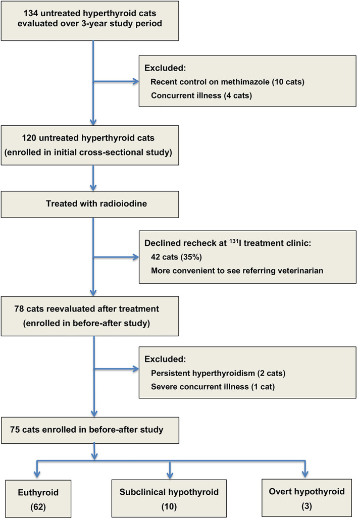
Animals: Forty-eight clinically normal cats and 120 cats with untreated hyperthyroidism, 75 of which were reevaluated after radioiodine-131 therapy.
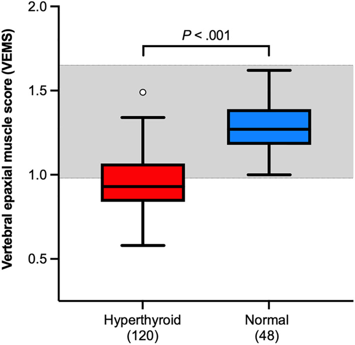
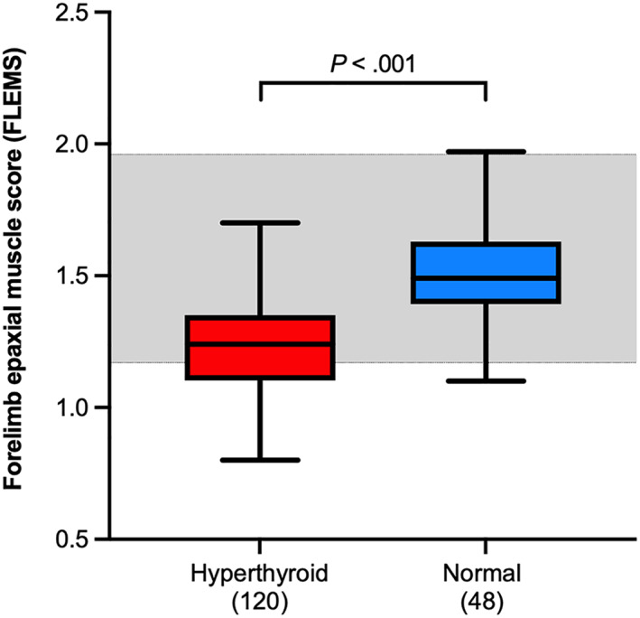
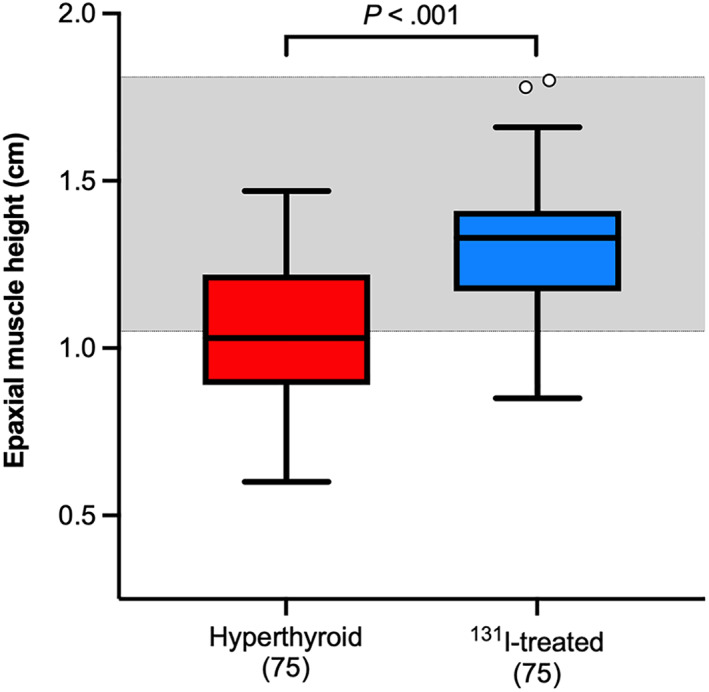
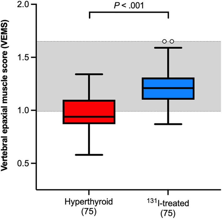
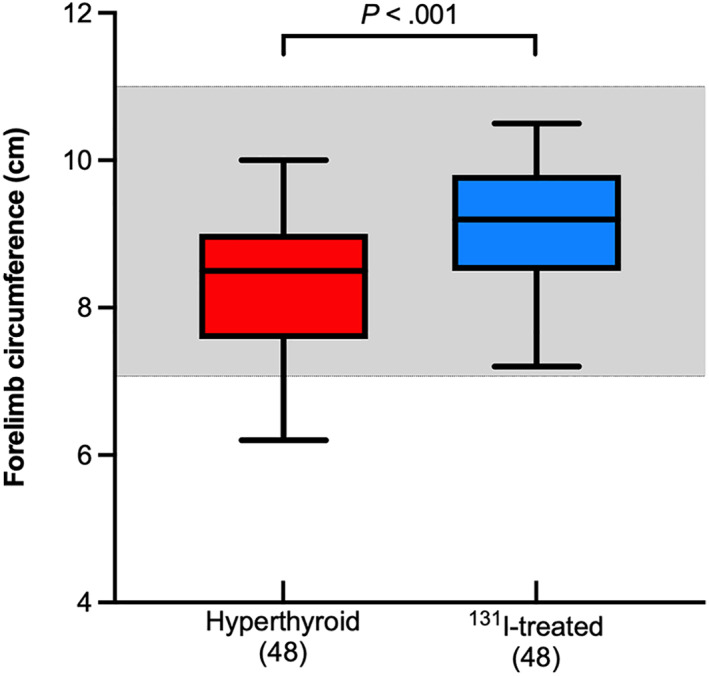
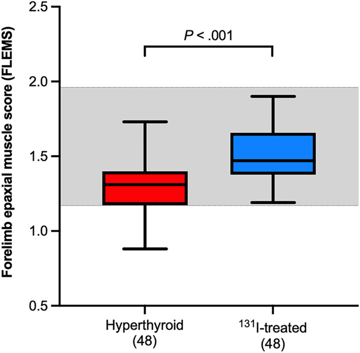
Methods: Prospective cross-sectional and before-after studies. All cats underwent ultrasonography and measurement of epaxial muscle height (EMH), with subsequent calculation of vertebral and forelimb epaxial muscle scores (VEMS and FLEMS). A subset of hyperthyroid cats underwent repeat muscle imaging 6 months after treatment.
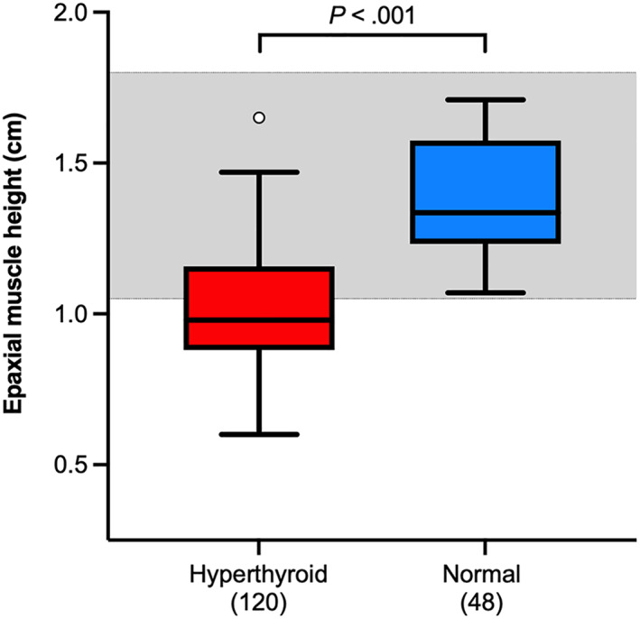
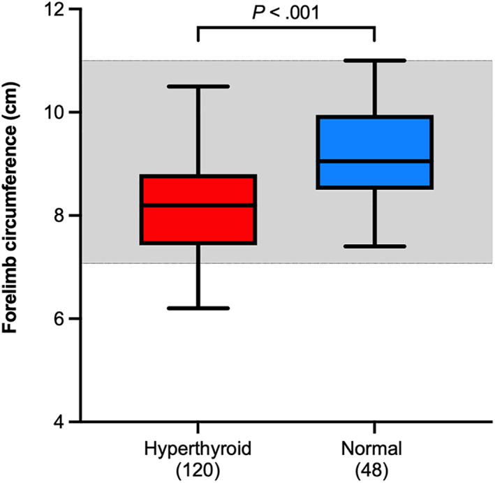
Results: Untreated hyperthyroid cats had a lower EMH than did clinically normal cats (median [25th-75th percentile], 0.98 [0.88-1.16] cm vs 1.34 [1.23-1.58] cm, P < .001). Seventy-seven (64.2%) untreated cats had subnormal EMH. Similarly, compared to normal cats, hyperthyroid cats had lower VEMS (0.93 [0.84-1.07] vs 1.27 [1.18-1.39], P < .001) and FLEMS (1.24 [1.10-1.35] vs 1.49 [1.39-1.63], P < .001). After treatment, EMH increased (1.03 [0.89-1.03] cm to 1.33 [1.17-1.41] cm, P < .001), with abnormally low EMH normalizing in 36/41 (88%). Both VEMS (0.94 [0.87-1.10] to 1.21 [1.10-1.31], P < .001) and FLEMS (1.31 [1.17-1.40] to 1.47 [1.38-1.66], P < .001) also increased after treatment.
Conclusions and clinical importance: Almost two-thirds of hyperthyroid cats have abnormally low muscle mass when measured quantitatively by ultrasound. Successful treatment restores muscle mass in >85% of cats. EMH provided the best means of quantitating muscle mass in these cats.
Keywords: 131I; cachexia; feline; muscle condition; radioactive iodine; sarcopenia; thyroid gland; ultrasound.
© 2022 The Authors. Journal of Veterinary Internal Medicine published by Wiley Periodicals LLC on behalf of American College of Veterinary Internal Medicine.
Conflict of interest statement
Authors declare no conflict of interest.
Figures
References
-
- Acotto CG, Niepomniszcze H, Mautalen CA. Estimating body fat and lean tissue distribution in hyperthyroidism by dual‐energy X‐ray absorptiometry. J Clin Densitom. 2002;5:305‐311. - PubMed
-
- Lonn L, Stenlof K, Ottosson M, et al. Body weight and body composition changes after treatment of hyperthyroidism. J Clin Endocrinol Metab. 1998;83:4269‐4273. - PubMed
-
- de la Rosa RE, Hennessey JV, Tucci JR. A longitudinal study of changes in body mass index and total body composition after radioiodine treatment for thyrotoxicosis. Thyroid. 1997;7:401‐405. - PubMed
-
- Da Nobrega AC, Vaisman M, De Araujo CG. Skeletal muscle function and body composition of patients with hyperthyroidism. Med Sci Sports Exerc. 1997;29:175‐180. - PubMed
MeSH terms
Substances
LinkOut - more resources
Full Text Sources
Medical
Miscellaneous
