. 2022 Oct 26;88(20):e0129922.
doi: 10.1128/aem.01299-22. Epub 2022 Oct 6.

Role of Bifidobacterium pseudocatenulatum in Degradation and Consumption of Xylan-Derived Carbohydrates

Affiliations

Role of Bifidobacterium pseudocatenulatum in Degradation and Consumption of Xylan-Derived Carbohydrates

Elizabeth Drey et al. Appl Environ Microbiol. .

Abstract

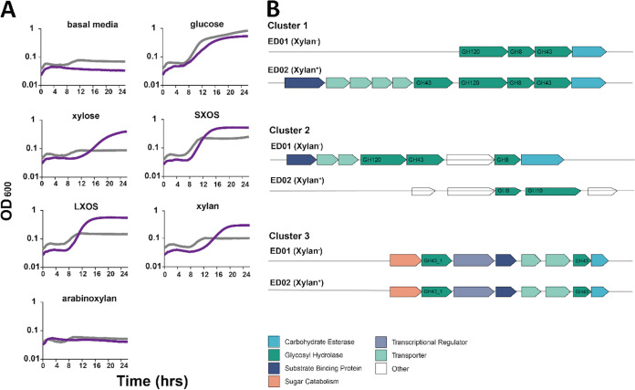
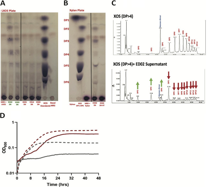
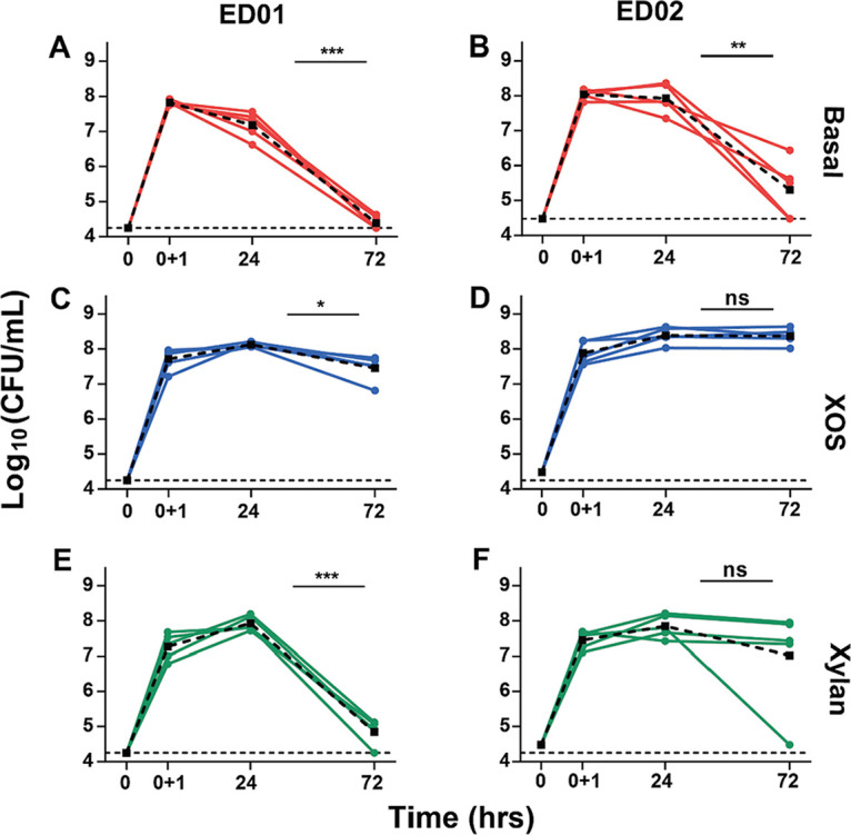
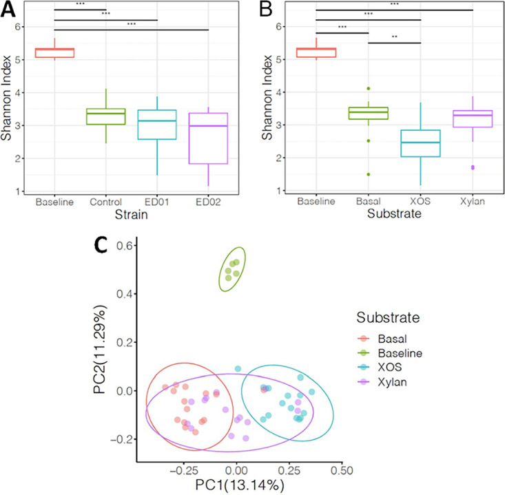
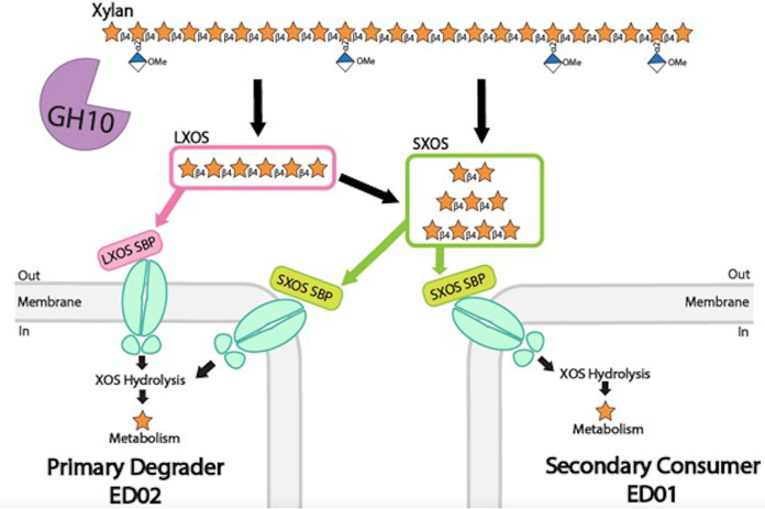
Xylans, a family of xylose-based polysaccharides, are dietary fibers resistant to digestion. They therefore reach the large intestine intact; there, they are utilized by members of the gut microbiota. They are initially broken down by primary degraders that utilize extracellular xylanases to cleave xylan into smaller oligomers. The resulting xylooligosaccharides (XOS) can either be further metabolized directly by primary degraders or cross-feed secondary consumers, including Bifidobacterium. While several Bifidobacterium species have metabolic systems for XOS, most grow poorly on longer-chain XOS and xylan substrates. In this study, we isolated strains of Bifidobacterium pseudocatenulatum and observed that some, including B. pseudocatenulatum ED02, displayed growth on XOS with a high degree of polymerization (DP) and straight-chain xylan, suggesting a primary degrader phenotype that is rare in Bifidobacterium. In silico analyses revealed that only the genomes of these xylan-fermenting (xylan+) strains contained an extracellular GH10 endo-β-1.4 xylanase, a key enzyme for primary degradation of xylan. The presence of an extracellular xylanase was confirmed by the appearance of xylan hydrolysis products in cell-free supernatants. Extracellular xylanolytic activity was only detected in xylan+ strains, as indicated by the production of XOS fragments with a DP of 2 to 6, identified by thin-layer chromatography (TLC) and high-performance liquid chromatography (HPLC). Additionally, in vitro fecal fermentations revealed that strains with a xylan+ phenotype can persist with xylan supplementation. These results indicate that xylan+ B. pseudocatenulatum strains may have a competitive advantage in the complex environment of the gastrointestinal tract, due to their ability to act as primary degraders of xylan through extracellular enzymatic degradation. IMPORTANCE The beneficial health effects of dietary fiber are now well established. Moreover, low fiber consumption is associated with increased risks of metabolic and systemic diseases. This so-called "fiber gap" also has a profound impact on the composition of the gut microbiome, leading to a disrupted or dysbiotic microbiota. Therefore, understanding the mechanisms by which keystone bacterial species in the gut utilize xylans and other dietary fibers may provide a basis for developing strategies to restore gut microbiome function. The results described here provide biochemical and genetic evidence for primary xylan utilization by human-derived Bifidobacterium pseudocatenulatum and show also that cooperative utilization of xylans occurs among other members of this species.

Keywords: bifidobacteria; cooperation; glycoside hydrolase; prebiotic; xylan; xylanase; xylooligosaccharide.


Conflict of interest statement

The authors declare a conflict of interest. R.H. has received grants and honoraria from several food and ingredient companies, is a co-owner of Synbiotic Health, and was on the Board of Directors of the International Scientific Association for Probiotics and Prebiotics.

Figures

FIG 1
FIG 1
Differences in growth phenotypes and XOS-active gene clusters between xylan+ ED02 and xylan ED01 B. pseudocatenulatum strains. (A) Growth of B. pseudocatenulatum xylan+ strain ED02 (purple) and xylan strain ED01 (gray) on various xylose-based carbohydrates. Cells were grown in modified deMan, Rogosa, and Sharpe (mMRS) medium supplemented with 0.5% carbohydrate. Basal medium, with no added carbohydrate, was used as a negative control, and glucose was used as a positive control. Other carbohydrate treatments included xylose, short-chain XOS (enriched in DP less than 4), long-chain XOS (enriched in DP greater than 4), xylan, and arabinoxylan. (B) Presence of XOS-active gene clusters between xylan+ and xylan phenotypes. Genes encoding XOS-active clusters I, II, and III are aligned according to their phenotype and are color-coded for relevant carbohydrate-active functions.
FIG 2
FIG 2
Production of endoxylanase and extracellular xylanase in B. pseudocatenulatum ED02 supports cross-feeding relationships between xylan+ and xylan strains. Shown are thin-layer chromatography (TLC) results of supernatant inoculated media containing either LXOS (lane 1, LXOS medium; lanes 2 to 6, LXOS media inoculated with respective strain supernatant; lane 7, 15 mM XOS standards DP1 to DP6; lane 8, basal medium) (A) or xylan (lane 1, xylose; lane 2, 15 mM XOS standards DP1 to DP6; lane 3, xylan medium; lane 4, xylan medium inoculated with ED02 supernatant; lane 5, xylan medium inoculated with heat-treated ED02 supernatant) (B). The chromatograms are composite of lanes from the same TLC plate. (C) Growth of xylan strain ED01 in the presence (red) or absence (gray) of ED02 supernatant-inoculated LXOS (dashed line) and xylan (solid line). (D) High-performance liquid chromatography (HPLC) indicates endoxylanase activity by ED02 producing small XOS fractions. Degradation products by ED02 supernatants after 48 h of incubation with XOS were separated and quantified by HPLC. In the XOS media, DP2 to -4 increased and DP5 to -13 decreased. Trends are indicated by arrows, displaying change in area under the curve from time zero to 48 h.
FIG 3
FIG 3
Test of persistence of B. pseudocatenulatum strains in fecal fermentations. Shown is qPCR quantification of strains ED01 and ED02 on basal medium (A and B), XOS (C and D), and xylan (E and F) in fecal fermentations with samples from 5 subjects. Persistence in individual fecal samples is shown as solid lines with colored circles, and averages are shown as dashed lines with square symbols. Significant differences between 24 h and 72 h were determined using a repeated-measure mixed model (*, P < 0.05; **, P < 0.01; ***, P < 0.001).
FIG 4
FIG 4
Analysis of microbial diversity and community composition through 16S rRNA sequencing of fecal fermentations at baseline and 72 h. (A and B) Shannon index was used to measure alpha diversity, and comparisons were made between strain (A) and substrate (B) treatments with baseline and respective controls. Significant differences are indicated by asterisks (Wilcoxon rank sum test with false-discovery rate [FDR] correction; *, P < 0.05; **, P < 0.01; ***, P < 0.001). (C) Beta diversity between substrates was visualized by principal-coordinate analysis (PCoA) based on Jaccard Index. Ellipses indicate 95% confidence intervals.
FIG 5
FIG 5
Model for xylan and XOS utilization by B. pseudocatenulatum. The xylan+ phenotype is implicated as a primary degrader due to the secretion of an extracellular xylanase that degrades xylan into smaller oligomers. The latter are available for transport by the primary degrader, or they can cross-feed other organisms. In contrast, strains having a xylan phenotype may still be secondary consumers. Such strains do not have the extracellular enzymatic machinery or transporters to consumer larger XOS and xylan molecules; they instead rely on other members of the microbial community to degrade xylans into smaller oligosaccharides that can serve as substrates for relevant transporters.

Similar articles

Cited by

References

    1. Bajpai P. 2014. Xylanolytic enzymes. Elsevier Inc, Amsterdam, The Netherlands.
    1. Faik A. 2010. Xylan biosynthesis: news from the grass. Plant Physiol 153:396–402. 10.1104/pp.110.154237. - DOI - PMC - PubMed
    1. Broxterman SE, Schols HA. 2018. Characterisation of pectin-xylan complexes in tomato primary plant cell walls. Carbohydr Polym 197:269–276. 10.1016/j.carbpol.2018.06.003. - DOI - PubMed
    1. Zhan P, Ye J, Lin X, Zhang F, Lin D, Zhang Y, Tang K. 2020. Complete genome sequence of Echinicola rosea JL3085, a xylan and pectin decomposer. Mar Genomics 52:100722. 10.1016/j.margen.2019.100722. - DOI - PubMed
    1. Jia X, Mi S, Wang J, Qiao W, Peng X, Han Y. 2014. Insight into glycoside hydrolases for debranched xylan degradation from extremely thermophilic bacterium Caldicellulosiruptor lactoaceticus. PLoS One 9:e106482. 10.1371/journal.pone.0106482. - DOI - PMC - PubMed

Publication types

LinkOut - more resources