. 2022 Oct 14;50(18):10503-10525.
doi: 10.1093/nar/gkac853.

APE1 assembles biomolecular condensates to promote the ATR-Chk1 DNA damage response in nucleolus

Affiliations

APE1 assembles biomolecular condensates to promote the ATR-Chk1 DNA damage response in nucleolus

Jia Li et al. Nucleic Acids Res. .

Abstract

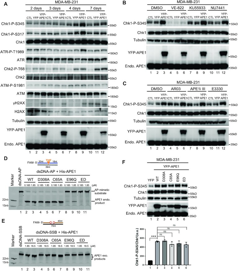
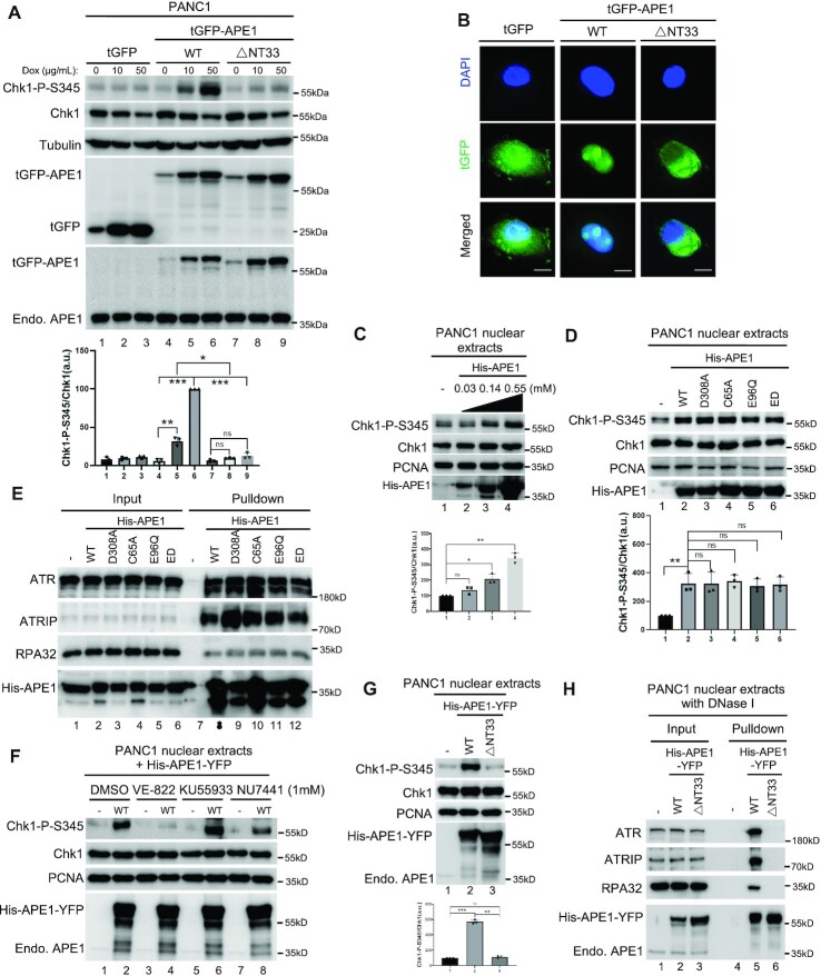
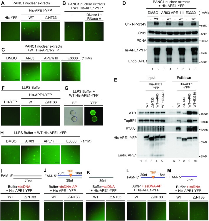
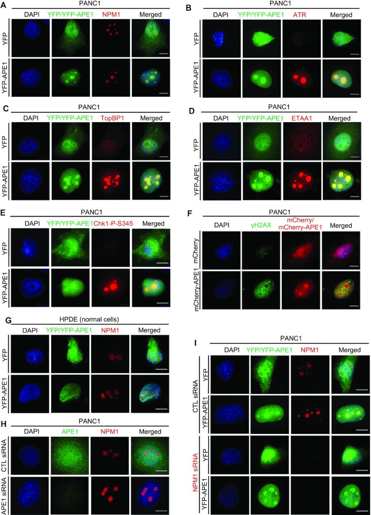
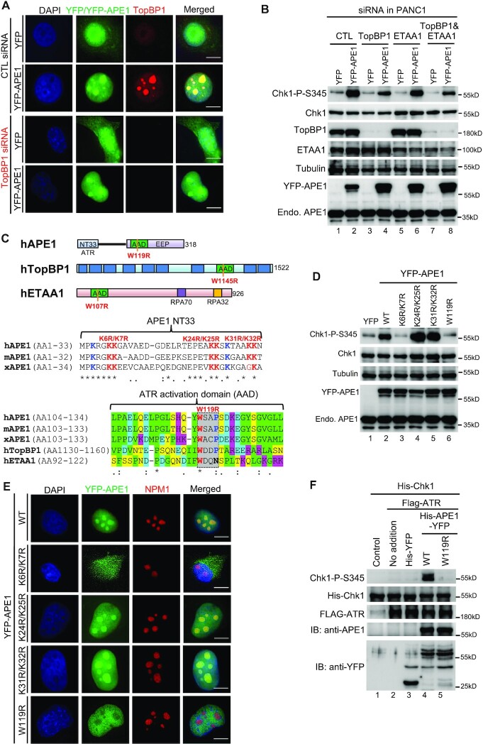
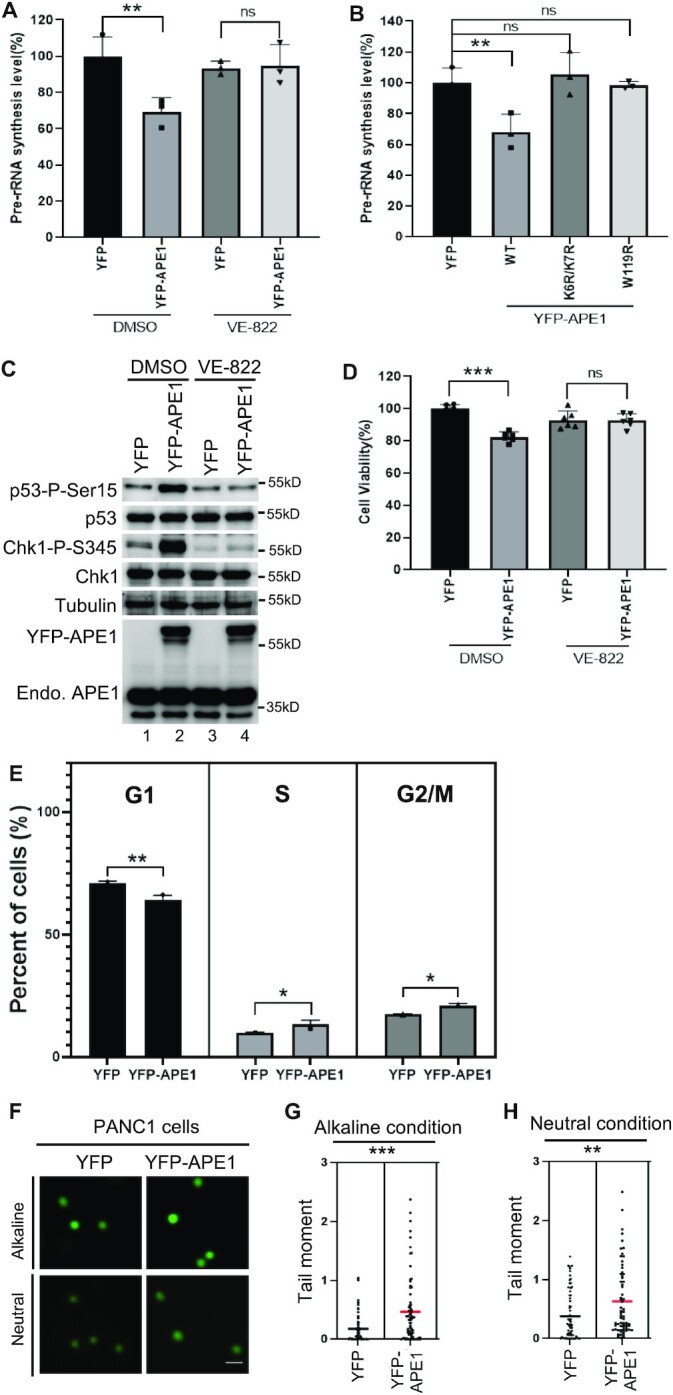
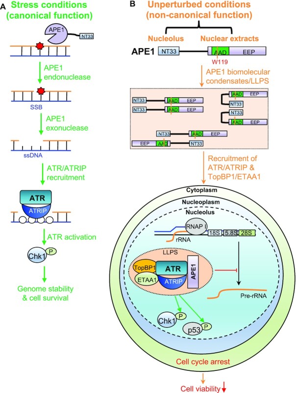
Multifunctional protein APE1/APEX1/HAP1/Ref-1 (designated as APE1) plays important roles in nuclease-mediated DNA repair and redox regulation in transcription. However, it is unclear how APE1 regulates the DNA damage response (DDR) pathways. Here we show that siRNA-mediated APE1-knockdown or APE1 inhibitor treatment attenuates the ATR-Chk1 DDR under stress conditions in multiple immortalized cell lines. Congruently, APE1 overexpression (APE1-OE) activates the ATR DDR under unperturbed conditions, which is independent of APE1 nuclease and redox functions. Structural and functional analysis reveals a direct requirement of the extreme N-terminal motif within APE1 in the assembly of distinct biomolecular condensates in vitro and DNA/RNA-independent activation of the ATR DDR. Overexpressed APE1 co-localizes with nucleolar NPM1 and assembles biomolecular condensates in nucleoli in cancer but not non-malignant cells, which recruits ATR and activator molecules TopBP1 and ETAA1. APE1 protein can directly activate ATR to phosphorylate its substrate Chk1 in in vitro kinase assays. W119R mutant of APE1 is deficient in nucleolar condensation, and is incapable of activating nucleolar ATR DDR in cells and ATR kinase in vitro. APE1-OE-induced nucleolar ATR DDR activation leads to compromised ribosomal RNA transcription and reduced cell viability. Taken together, we propose distinct mechanisms by which APE1 regulates ATR DDR pathways.


Figures

Figure 1.
Figure 1.
APE1 and its nuclease activity is important for ATR–Chk1 DDR pathway in mammalian cells. (A) siRNA-mediated APE1-knockdown impaired H2O2-induced ATR–Chk1 DDR pathway in MDA-MB-231 cells. Total cell lysates were extracted and analyzed via immunoblotting analysis as indicated. (B, C) The H2O2-induced Chk1 phosphorylation was compromised by AR03 or APE1i III at different doses (5, 10, or 15 μM for 2 h) in MDA-MB-231 cells. (D) The H2O2-induced Chk1 phosphorylation was reduced in APE1-KD PANC1 cells. (E, F) The H2O2-induced Chk1 phosphorylation was impaired by AR03 or APE1i III in a dose-dependent manner (5, 10 or 15 μM for 2 h) in PANC1 cells. (A–F) ‘Chk1-P-S345/Chk1’ was quantified by the intensity of Chk1-P-S345 versus total Chk1. Means ± SD, n = 3. (G) AR03 or APE1i III but not E3330 inhibited APE1’s endonuclease activity. Different doses of AR03, APE1i III, or E3330 (1, 2, or 5 mM) was added to endonuclease assays containing 0.185 μM APE1 protein and a 5’-FAM-labeled dsDNA-AP substrate. After incubation for 30 min, samples were analyzed via 20% TBE gel. (H) AR03 or Inhibitor III could inhibit APE1’s exo-nuclease activity. Different doses of AR03, APE1i III, or E3330 (1, 2 or 5 mM) was added to exonuclease assays containing 0.185 μM APE1 protein and 5’-FAM-labeled dsDNA-SSB substrate. After 30-min incubation, samples were examined via 20% TBE gel.
Figure 2.
Figure 2.
APE1 overexpression leads to the activation of ATR–Chk1 DDR pathway independent of its nuclease and redox function in mammalian cells. (A) ATR–Chk1 DDR pathway was activated by the overexpression of YFP-APE1 but not YFP nor CTL transfection (no vector transfection) in MDA-MB-231 cells. After different times of incubation (i.e. 2, 3, 4 or 7 days), total cell lysates were extracted and examined via immunoblotting analysis as indicated. (B) Chk1 phosphorylation induced by YFP-APE1 was dependent on ATR, but not ATM nor DNA-PKcs. After 4-d overexpression of YFP-APE1 or YFP, different DDR kinase inhibitors (5 μM of VE-822, KU55933 or NU7441) were added to MDA-MB-231 cells for 2 h, followed by total cell lysate extraction and immunoblotting analysis as indicated. (C) APE1 inhibitors almost had no noticeable effect on Chk1 phosphorylation by YFP-APE1 overexpression. After 2-day overexpression of YFP-APE1 or YFP, different APE1 inhibitors (1 μM of AR03, APE1i III or E3330) were added to MDA-MB-231 cells for 2 days, followed by total cell lysate extraction and immunoblotting analysis. (D) The endonuclease activity of WT or mutant (i.e. D308A, C64A, E96Q, ED) His-APE1 was examined on 20% TBE gel. (E) The exonuclease activity of WT or mutant (i.e. D308A, C64A, E96Q, ED) His-APE1 was examined on 20% TBE gel. (F) Chk1 phosphorylation was triggered by WT/mutant YFP-APE1 (i.e. D308A, C64A, E96Q, ED) overexpression in MDA-MB-231 cells. After 4-day overexpression of WT/mutant YFP-APE1 or YFP in MDA-MB-231, total cell lysates were extracted and analyzed via immunoblotting analysis. The Chk1-P-S345/Chk1 data are presented as means ± SD, n = 3.
Figure 3.
Figure 3.
The APE1 N33 motif is required for ATR–Chk1 DDR pathway in vivo and in vitro. (A) Overexpression of WT but not △N33 tGFP-APE1 nor tGFP activated Chk1 phosphorylation in PANC1 cells. APE1 or N33-depletion over-expression plasmid was transfected to PANC1 cells. After 1-day incubation of various overexpression plasmid transfection, different doses of doxycycline (0, 10 or 50 μg/ml) were added to PANC1 cells for another 2 days. Total cell lysates were then extracted and analyzed via immunoblotting analysis. (B) △N33 tGFP-APE1 was deficient in nuclear localization in PANC1 cells. tGFP, WT or △N33 tGFP-APE1 overexpression plasmid was transfected to PANC1 cells. After 1-day incubation of overexpression plasmid transfection, 50 μg/ml doxycycline was added to PANC1 cells for another 2 days, followed with fluorescence microscopy analysis. Scale bars = 10 μm. (C) Chk1 phosphorylation was triggered by the excess addition of recombinant His-APE1 protein in a dose-dependent manner in PANC1 nuclear extracts. Different doses of His-APE1 protein were added to PANC1 nuclear extracts for incubation for 30 min at room temperature. The samples were analyzed via immunoblotting analysis as indicated. (D) Chk1 phosphorylation induced by excess addition of recombinant His-APE1 protein in PANC1 nuclear extracts was independent of its nuclease activity and redox function. WT or mutant (i.e. D308A, C65A, E96Q, ED) His-APE1 was added to PANC1 nuclear extracts for 30-min incubation at room temperature, followed by immunoblotting analysis. (E) Pulldown assays suggest that WT/mutant His-APE1 associated with ATR, ATRIP and RPA32 in PANC1 nuclear extracts. ‘Input’ or ‘Pulldown’ samples were examined via immunoblotting analysis as indicated. (F) Chk1 phosphorylation induced by WT His-APE1-YFP protein in PANC1 nuclear extracts was inhibited by VE-822 but not KU55933 nor NU7441 (1 mM). (G) Chk1 phosphorylation was activated by excess addition of WT His-APE1-YFP but not △N33 His-APE1-YFP protein in PANC1 nuclear extracts. (H) Pulldown assays suggest that WT but not △N33 His-APE1-APE1 associated with ATR, ATRIP and RPA32 in PANC1 nuclear extracts pre-treated with DNase I (2 mg/ml). ‘Input’ or ‘Pulldown’ samples were examined via immunoblotting analysis as indicated. (A, C, D, G) Chk1-P-S345/Chk1 was quantified and examined as means ± SD (n = 3).
Figure 4.
Figure 4.
APE1 assembles biomolecular condensates in vitro to promote ATR DDR pathway. (A) WT but not △N33 His-APE1-YFP formed phase separation in PANC1 nuclear extracts. Recombinant YFP, WT or △N33 His-APE1-YFP protein (0.55 mM) was added to PANC1 nuclear extract and incubated for 15 min at room temperature, followed by fluorescence microscopy analysis. (B) DNase I and RNase A had almost no effect on the APE1-assembled phase separation in nuclear extracts. DNase I and RNase A (2 mg/ml each) was added to PANC1 nuclear extracts and incubated for 30 min at 37°C. Then WT His-APE1-YFP protein (0.55 mM) was added and incubated for 10 min at room temperature, followed by fluorescence microscopy analysis. (C) APE1 inhibitors had no noticeable effect on phase separation formed by APE1 in nuclear extracts. WT His-APE1-YFP protein (0.55 mM) was added to PANC1 supplemented with DMSO or APE1 inhibitors (AR03, APE1i III, and E3330). After 15-min incubation at room temperature, reaction mixtures were examined via fluorescence microscopy analysis. (D) APE1 inhibitors had no effect on the Chk1 phosphorylation induced by the excess addition of APE1 protein in PANC1 nuclear extracts. APE1 inhibitor (AR03, APE1i III or E3330) was added to PANC1 nuclear extracts and incubated for 10 min at room temperature, which was supplemented with WT His-APE1-YFP (0.55 mM) and incubated for another 30 min at room temperature. The samples were then analyzed via immunoblotting analysis as indicated. (E) WT but not △N33 His-APE1-YFP associated with ATR, TopBP1 and ETAA1 in PANC1 nuclear extracts. ‘Input’ and ‘Pulldown’ samples from pulldown experiments were examined via immunoblotting analysis as indicated. AR03 or E3330 was added to 1 mM. (F) WT but not △N33 His-APE1-YFP formed phase separation in a LLPS buffer. WT but not △N33 His-APE1-YFP (0.55 mM) was added to a LLPS buffer and incubated for 15 min at room temperature, followed by fluorescence microscopy analysis. (G) Phase separation formed by recombinant WT His-APE1-YFP protein was fused into large biomolecular condensates in a LLPS buffer. WT His-APE1-YFP (0.55 mM) was added to a buffer and incubated for 2 h before fluorescence microscopy analysis. BF, bright field. (H) Phase separation assembled by APE1 in a LLPS buffer was not affected by the APE1 inhibitors. WT His-APE1-YFP protein (0.55 mM) was added to LLPS buffer supplemented with DMSO or APE1 inhibitors (AR03, APE1i III and E3330). After 15-min incubation at room temperature, reaction mixtures were examined via fluorescence microscopy analysis. (I–M) The requirement of NT33 motif within APE1 for phase separation in a LLPS buffer was bypassed with the presence of different DNA or RNA. WT but not △N33 His-APE1-YFP (0.55 mM) was added to a buffer supplemented with 0.5 μM of different DNA/RNA structures (dsDNA (I), dsDNA with AP site (J), ssDNA (K), ssDNA with AP site (L) and ssRNA (M)). Reaction mixtures were incubated for 15 min at room temperature before fluorescence microscopy analysis. All scale bars = 50 μm.
Figure 5.
Figure 5.
APE1 forms biomolecular condensates in nucleoli to recruit ATR, TopBP1, and ETAA1 for ATR activation in a NPM1-independent fashion in cancer cells but not unmalignant cells. (A) Overexpressed YFP-APE1 but not YFP colocalized with NPM1 in nucleoli in PANC1 cells. After overexpression of YFP or YFP-APE1 for 3 days, PANC1 cells were fixed and incubated with anti-NPM1-AF647 fluorescence antibody for overnight at 4°C. (B–D) ATR and its activators TopBP1 and ETAA1 were colocalized with YFP-APE1 in nucleoli in PANC1 cells. YFP or YFP-APE1 overexpression plasmid was added to PANC1 cells. After overexpression of YFP or YFP-APE1 for 3 days, PANC1 cells were fixed and incubated with anti-ATR-AF647 (B), anti-TopBP1-AF647 (C), or anti-ETAA1-AF647 (D) for overnight at 4°C. (E) After overexpression of YFP or YFP-APE1 for 3 days, PANC1 cells were fixed and incubated with anti-Chk1-P-S345 antibody for overnight at 4°C, followed by incubation with anti-Rabbit secondary antibody conjugated with AF594 at 4°C for 4 h. (F) After overexpression of mCherry or mCherry-APE1 for 3 days, PANC1 cells were fixed and incubated with anti-γH2AX-AF488 at 4°C overnight. (G) Overexpressed YFP-APE1 was not colocalized with NPM1 in nucleoli in HPDE cells. Similar experiment to Panel (A) was performed in HPDE cells. (H) Nucleolar localization of NPM1 was not affected when endogenous APE1 was knocked down via siRNA. Control (CTL) siRNA or APE1 siRNA was added and transfected to PANC1 cells for 3 days. The cells were then fixed and incubated with anti-APE1-AF488 and anti-NPM1-AF647 for overnight at 4°C. (I) YFP-APE1 still assembled condensates when endogenous NPM1 was knocked down. CTL siRNA or NPM1 siRNA was transfected to PANC1 cells and incubated for 1 day. Then YFP or YFP-APE1 overexpression plasmid was transfected and incubated for 3 days. The cells were fixed and incubated with anti-NPM1-AF647 at 4°C overnight. (A–I) All cells were examined by fluorescence microscopy analysis. All scale bars = 10 μm.
Figure 6.
Figure 6.
While TopBP1 is dispensable for YFP-APE1 nucleolar localization, the W119 residue within APE1’s putative ATR activation domain is required for its biomolecular condensate formation in nucleoli and nucleolar ATR activation. (A) YFP-APE1 still assembled condensates and activated ATR-chk1 pathway when endogenous TopBP1 was knocked down via siRNA. After 1-d incubation of CTL or TopBP1 siRNA in PANC1 cells, YFP or YFP-APE1 overexpression plasmid was added and incubation for another 3 d. The cells were fixed and incubated with anti-TopBP1-AF647 for fluorescence microscopy analysis. (B). After 1-d incubation of CTL, TopBP1 and/or ETAA1 siRNA in PANC1 cells, YFP or YFP-APE1 overexpression plasmid was added and incubated for another 3 d. The cells were extracted and examined by immunoblotting analysis as indicated. (C) Schematic diagrams of functional domains of hAPE1, hTopBP1 and hETAA1 as well as sequence alignment of the N-terminal domain and putative ATR activation domain (AAD) within APE1. hAPE1 (human APE1, NCBI#: P27695.1), mAPE1 (mouse APE1, NCBI#: NP_033817.1), xAPE1 (Xenopus laevis APE1, NCBI#: AAH72056.1), hTopBP1 (human TopBP1, NCBI#: Q92547.3), and hETAA1 (human ETAA1, NCBI#: NP_061875.2). * identical;: highly conserved;. low conservation. In the AAD alignment, highlighted green indicates aa with hydrophobic side chains, highlighted yellow indicates aa with polar uncharged side chains, highlighted pink indicates aa with positive charged sides chains, highlighted turquoise indicates aa with negative charged sides chains, and highlighted gray indicates the conserved WxxP/N peptide. (D) The K6, K7 and W119 residues within APE1 are important for Chk1 phosphorylation induced by YFP-APE1 in PANC1 cells. After YFP, WT or mutant (i.e. K6R/K7R, K24R/K25R, K31R/K32R, W119R) YFP-APE1 overexpression plasmid was added to PANC1 cells and incubation for 3 d, total cell lysates were extracted and examined via immunoblotting analysis as indicated. (E) APE1 forms biomolecular condensates in nucleoli dependent on its W119 residue. After WT or mutant (i.e. K6R/K7R, K24R/K25R, K31R/K32R, W119R) YFP-APE1 overexpression plasmid was added to PANC1 cells and incubation for 3 d, the cells were fixed and incubated with anti-NPM1-AF647 for overnight at 4°C, followed by fluorescence microscopy analysis. (F) WT but not W119R YFP-APE1 directly activates ATR to phosphorylate Chk1 phosphorylaton at S345 in vitro. In in vitro kinase assays, equal amount of His-YFP, WT or W119R His-APE1-YFP was added to the kinase assays (purified Flag-ATR as kinase and purified His-Chk1 as substrate). Beads bound with lysates from HEK293 cells without Flag-ATR transfection were used as a negative control (‘Control’). ‘No addition’ indicate the kinase assays without the addition of His-YFP or WT/W119R His-APE1-YFP. The samples were examined via immunoblotting analysis as indicated. (A, E) All scale bars = 10 μm.
Figure 7.
Figure 7.
APE1-induced ATR–Chk1 nDDR leads to rRNA transcription suppression and cell viability reduction. (A) APE1 overexpression led to pre-rRNA transcription suppression in an ATR-dependent fashion. YFP or YFP-APE1 overexpression plasmid was added to PANC1 cells and incubated for 2 days. DMSO or VE-822 (1 μM) was then added and incubated for another day. The total RNA was extracted and analyzed via qRT-PCR, and pre-rRNA synthesis was normalized to beta-Actin. n = 3. (B) WT but not K6R/K7R nor W119R YFP-APE1 suppressed rRNA transcription. YFP or WT or mutant YFP-APE1 overexpression plasmid was added to PANC1 cells and incubated for 3 days, followed by total RNA extraction and qRT-PCR analysis. n = 3. (C) Overexpression of YFP-APE1 but not YFP led to p53 Ser15 phosphorylation, which was sensitive to VE-822. After YFP or YFP-APE1 overexpression plasmid was added to PANC1 cells and incubated for 3 days, DMSO or VE-822 (1 μM) was added and incubated for another 2 h, followed by total cell lysate extraction and immunoblotting analysis as indicated. (D) YFP-APE1 overexpression led to cell viability reduction. After YFP or YFP-APE1 overexpression plasmid was added to PANC1 cells and incubated for 2 days, DMSO or VE-822 (1 μM) was added and incubated for another day. The cells were analyzed via MTT assays. n = 6. (E) Overexpression of YFP-APE1 led to cell cycle arrests at S and G2/M phases in PANC1 cells. YFP or WT YFP-APE1 overexpression plasmid was added to PANC1 cells. After incubation for 4 days, the cells were collected and examined via FACS analysis. The data are presented as means ± SD, n = 3. (F–H) Overexpression APE1 led to more endogenous DNA damage in PANC1 cells. YFP or WT YFP-APE1 overexpression plasmid was added to PANC1 cells. After incubation for 4 days, the cells were collected and analyzed via comet assays under alkaline or neutral conditions. Scale bar = 100 μm. The data are presented as Scatter dot plot and the lines indicate means, n > 50.
Figure 8.
Figure 8.
A working model of distinct mechanisms of APE1 in the ATR DNA damage response. (A) Stress conditions (canonical function): APE1 promotes the ATR–Chk1 DDR pathway activation via its AP endonuclease and exonuclease activities under stress conditions, which is considered as canonical function of APE1. (B) Unperturbed conditions (non-canonical function): under unperturbed conditions, APE1 overexpression leads to biomolecular condensates (liquid–liquid phase separation) in nucleoli in vivo and in nuclear extracts in vitro via its N33 motif and/or W119 residue in AAD domain. ATR/ATRIP, TopBP1, and ETAA1 are recruited to the APE1-induced condensates for ATR activation. APE1-induced nucleolar ATR DDR suppresses rRNA transcription, arrests cell cycle, elevates DNA damage, and reduces cell viability. See text for more details.

Similar articles

Cited by

References

    1. Tubbs A., Nussenzweig A.. Endogenous DNA damage as a source of genomic instability in cancer. Cell. 2017; 168:644–656. - PMC - PubMed
    1. Ciccia A., Elledge S.J.. The DNA damage response: making it safe to play with knives. Mol. Cell. 2010; 40:179–204. - PMC - PubMed
    1. Lindahl T., Barnes D.E.. Repair of endogenous DNA damage. Cold Spring Harb. Symp. Quant. Biol. 2000; 65:127–133. - PubMed
    1. Lindahl T. Instability and decay of the primary structure of DNA. Nature. 1993; 362:709–715. - PubMed
    1. Thompson P.S., Cortez D. New insights into abasic site repair and tolerance. DNA Repair (Amst.). 2020; 90:102866. - PMC - PubMed

Publication types

MeSH terms