Cerebellar Functional Dysconnectivity in Drug-Naïve Patients With First-Episode Schizophrenia
- PMID: 36200880
- PMCID: PMC10016395
- DOI: 10.1093/schbul/sbac121
Cerebellar Functional Dysconnectivity in Drug-Naïve Patients With First-Episode Schizophrenia
Abstract
Background: Cerebellar functional dysconnectivity has long been implicated in schizophrenia. However, the detailed dysconnectivity pattern and its underlying biological mechanisms have not been well-charted. This study aimed to conduct an in-depth characterization of cerebellar dysconnectivity maps in early schizophrenia.
Study design: Resting-state fMRI data were processed from 196 drug-naïve patients with first-episode schizophrenia and 167 demographically matched healthy controls. The cerebellum was parcellated into nine functional systems based on a state-of-the-art atlas, and seed-based connectivity for each cerebellar system was examined. The observed connectivity alterations were further associated with schizophrenia risk gene expressions using data from the Allen Human Brain Atlas.
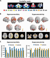
Study results: Overall, we observed significantly increased cerebellar connectivity with the sensorimotor cortex, default-mode regions, ventral part of visual cortex, insula, and striatum. In contrast, decreased connectivity was shown chiefly within the cerebellum, and between the cerebellum and the lateral prefrontal cortex, temporal lobe, and dorsal visual areas. Such dysconnectivity pattern was statistically similar across seeds, with no significant group by seed interactions identified. Moreover, connectivity strengths of hypoconnected but not hyperconnected regions were significantly correlated with schizophrenia risk gene expressions, suggesting potential genetic underpinnings for the observed hypoconnectivity.
Conclusions: These findings suggest a common bidirectional dysconnectivity pattern across different cerebellar subsystems, and imply that such bidirectional alterations may relate to different biological mechanisms.
Keywords: cerebellum; genetic risk; hyperconnectivity; hypoconnectivity; schizophrenia.
© The Author(s) 2022. Published by Oxford University Press on behalf of the Maryland Psychiatric Research Center. All rights reserved. For permissions, please email: journals.permissions@oup.com.
Figures


References
-
- Ding Y, Ou Y, Pan P, et al. Cerebellar structural and functional abnormalities in first-episode and drug-naive patients with schizophrenia: a meta-analysis. Psychiatry Res Neuroimag. 2019;283:24–33. - PubMed
-
- Andreasen NC, Paradiso S, O’Leary DS.. “Cognitive dysmetria” as an integrative theory of schizophrenia: a dysfunction in cortical-subcortical-cerebellar circuitry? Schizophr Bull. 1998;24(2):203–218. - PubMed
-
- Guo W, Zhang F, Liu F, et al. Cerebellar abnormalities in first-episode, drug-naive schizophrenia at rest. Psychiatry Res Neuroimag. 2018;276:73–79. - PubMed
Publication types
MeSH terms
LinkOut - more resources
Full Text Sources
Medical

