. 2022 Dec 1;323(6):F633-F641.
doi: 10.1152/ajprenal.00107.2022. Epub 2022 Oct 6.

NOXA1-dependent NADPH oxidase 1 signaling mediates angiotensin II activation of the epithelial sodium channel

Affiliations

NOXA1-dependent NADPH oxidase 1 signaling mediates angiotensin II activation of the epithelial sodium channel

Elena Mironova et al. Am J Physiol Renal Physiol. .

Abstract

The activity of the epithelial Na+ channel (ENaC) in principal cells of the distal nephron fine-tunes renal Na+ excretion. The renin-angiotensin-aldosterone system modulates ENaC activity to control blood pressure, in part, by influencing Na+ excretion. NADPH oxidase activator 1-dependent NADPH oxidase 1 (NOXA1/NOX1) signaling may play a key role in angiotensin II (ANG II)-dependent activation of ENaC. The present study aimed to explore the role of NOXA1/NOX1 signaling in ANG II-dependent activation of ENaC in renal principal cells. Patch-clamp electrophysiology and principal cell-specific Noxa1 knockout (PC-Noxa1 KO) mice were used to determine the role of NOXA1/NOX1 signaling in ANG II-dependent activation of ENaC. The activity of ENaC in the luminal plasma membrane of principal cells was quantified in freshly isolated split-opened tubules using voltage-clamp electrophysiology. ANG II significantly increased ENaC activity. This effect was robust and observed in response to both acute (40 min) and more chronic (48-72 h) ANG II treatment of isolated tubules and mice, respectively. Inhibition of ANG II type 1 receptors with losartan abolished ANG II-dependent stimulation of ENaC. Similarly, treatment with ML171, a specific inhibitor of NOX1, abolished stimulation of ENaC by ANG II. Treatment with ANG II failed to increase ENaC activity in principal cells in tubules isolated from the PC-Noxa1 KO mouse. Tubules from wild-type littermate controls, though, retained their ability to respond to ANG II with an increase in ENaC activity. These results indicate that NOXA1/NOX1 signaling mediates ANG II stimulation of ENaC in renal principal cells. As such, NOXA1/NOX1 signaling in the distal nephron plays a central role in Na+ homeostasis and control of blood pressure, particularly as it relates to regulation by the renin-ANG II axis.NEW & NOTEWORTHY Activity of the epithelial Na+ channel (ENaC) in the distal nephron fine-tunes renal Na+ excretion. Angiotensin II (ANG II) has been reported to enhance ENaC activity. Emerging evidence suggests that NADPH oxidase (NOX) signaling plays an important role in the stimulation of ENaC by ANG II in principal cells. The present findings indicate that NOX activator 1/NOX1 signaling mediates ANG II stimulation of ENaC in renal principal cells.

Keywords: collecting duct; hypertension; reactive oxygen species; renal physiology; sodium excretion.


Conflict of interest statement

M.S.R. is a member of the Board of Directors at Eli Lilly & Company. None of the other authors has any conflicts of interest, financial or otherwise, to disclose.

Figures

None
Graphical abstract
Figure 1.
Figure 1.
Genotype of the principal cell-specific NADPH oxidase activator 1 knockout (PC-Noxa1 KO) mouse. Shown is an inverted image of representative gels showing typical PCR products from genotyping reactions with genomic DNA from littermate controls and PC-Noxa1 KO mice. Arrowheads indicate expected products of aquaporin 2 (Aqp2)-Cre and floxed Noxa1 transgenes as well as the Noxa1 allele that has undergone recombination (Noxa1del1). Genomic DNA was extracted from the renal medulla and cortex as well as the aorta. To maximize clarity, the gray scale of this image was inverted for presentation purposes.
Figure 2.
Figure 2.
Angiotensin II (ANG II) increases epithelial Na+ channel (ENaC) activity in principal cells of the connecting tubule (CNT)/cortical collecting duct (CCD). A: representative current traces of ENaC in cell-attached patches on apical plasma membranes of principal cells in the split-open CNT/CCD from wild-type C57BL/6J mice. Tubules were treated with vehicle (top) or 500 nM ANG II for 40 min (middle) or were from mice treated with 500 ng/kg/min ANG II (osmotic minipump) for 48-72 h (bottom). Summary graphs show means ± SE (bars) and individual data points (circles, boxes, and triangles; solid symbols indicate males and open symbols indicate females) for ENaC activity (NPo; B), open probability (Po; C), and number of active channels per patch (N; D) in tubules from the vehicle control (con; n = 21 cells from 3 male and 3 female mice), 40-min ANG II treatment (n = 11 cells from 3 male mice), and 48- to 72-h ANG II treatment (n = 10 cells from 2 females) groups. *Significantly different compared with the control by one-way ANOVA with a Tukey highly significant difference post hoc test. ns, not significant. C and O indicate closed and open channels, respectively.
Figure 3.
Figure 3.
Angiotensin II (ANG II) type 1 receptor blockade abolishes ANG II effects on the epithelial Na+ channel (ENaC). A: representative current traces of ENaC in cell-attached patches on apical plasma membranes of principal cells in the split-open connecting tubule/cortical collecting duct from wild type-C57BL/6J mice treated with 500 nM ANG II (top) or 500 nM ANG II plus 500 nM losartan for 40 min (bottom). The ANG II trace is identical to that shown in Fig. 2A and is reshown here for presentation purposes. Summary graphs showing means ± SE (bars) and individual data points (circles, boxes, and triangles; solid symbols indicate males and open symbols indicate females) for ENaC activity (NPo; B), open probability (Po; C), and number of active channels per patch (N; D) in tubules from the vehicle control (con; n = 21 cells from 3 male and female mice), 40-min ANG II treatment (n = 11 cells from 3 male mice), and 40-min ANG II plus losartan treatment (n = 10 cells from 2 male and 1 female mice) groups. The vehicle and ANG II treatment groups are identical those shown in Fig. 2, B–D, and are reshown here for presentation purposes. Significantly different compared with *control and **ANG II treatment groups by one-way ANOVA with a Tukey highly significant difference post hoc test. ns, not significant. C and O indicate closed and open channels, respectively.
Figure 4.
Figure 4.
Inhibition of NADPH oxidase 1 (NOX1) prevents epithelial Na+ channel (ENaC) activation by angiotensin (ANG II). A: representative current traces of ENaC after treatment with 500 nM ANG II for 40 min in the absence (top) and presence of 500 nM ML171 (bottom). The ANG II trace is identical to that shown in Fig. 2 and is reshown here for presentation purposes. Summary graphs showing means ± SE (bars) and individual data points (circles, boxes, and triangles; solid symbols indicate males and open symbols indicate females) for ENaC activity (NPo; B), open probability (Po; C), and number of active channels per patch (N; D) in tubules from the vehicle control (con; n = 21 cells from 3 male and 3 female mice), 40-min ML171 treatment (n = 13 cells from 2 male and 2 female mice), 40-min ANG II treatment (n = 11 cells from 3 male mice), and 40-min ANG II plus ML171 treatment (n = 12 from 3 male mice) groups. The vehicle and ANG II treatment groups are identical to those shown in Fig. 2. Significantly different compared with the *control and **ANG II treatment groups by one-way ANOVA with a Tukey highly significant difference post hoc test. ns, not significant. C and O indicate closed and open channels, respectively.
Figure 5.
Figure 5.
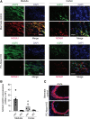
NADPH oxidase activator 1 (Noxa1) is specifically deleted in renal principal cells of the principal cell-specific Noxa1 knockout (PC-Noxa1 KO) mouse. A: representative fluorescence microscopy images of frozen renal medulla and cortex sections from littermate controls (top) and PC-Noxa1 KO (bottom) mice stained for immunoreactive NOXA1 (red), aquaporin 2 (Aqp2; green), and DAPI (blue). Scale bars = 100 µm. B: quantification of NOXA1 colocalization with AQP2 in the renal medulla and cortex. Data are fluorescence integrated density of merged fluorescence images (means ± SE, n = 5). C: representative fluorescence microscopy images of the frozen aorta from littermate controls (top) and PC-Noxa1 KO (bottom) mice stained for immunoreactive NOXA1 (red) and DAPI (blue). Scale bar = 100 μm.
Figure 6.
Figure 6.
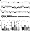
Angiotensin II (ANG II) had no effect on epithelial Na+ channel (ENaC) activity in principal cells of the principal cell-specific Noxa1 knockout (PC-Noxa1 KO) mouse. Representative current traces of ENaC in cell-attached patches on the apical plasma membranes of principal cells in the split-open connecting tubule/cortical collecting duct from littermate controls (A) and PC-Noxa1 KO mice (B) treated with vehicle (top) or 500 nM ANG II for 40 min (bottom). Summary graphs of means ± SE (bars) and individual data points for ENaC activity (NPo; C), open probability (Po; D), and number of channels (N; E) in principal cells from littermate controls (con; n = 13 cells from 2 male and 1 female mice, circles) without and with ANG II treatment (n = 15 cells from 2 male and 1 female mice, squares) and from PC-Noxa1 KO mice (n = 13 cells from 1 male and 3 female mice, triangles) without and with ANG II (n = 13 cells from 2 male and 2 female mice, diamonds). These experiments included mice of both sexes at approximately equal proportions. *Significantly different by one-way ANOVA with a Tukey highly significant difference post hoc test. ns, not significant. C and O indicate closed and open channels, respectively.

References

    1. Lifton RP, Wilson FH, Choate KA, Geller DS. Salt and blood pressure: new insight from human genetic studies. Cold Spring Harb Symp Quant Biol 67: 445–450, 2002. doi:10.1101/sqb.2002.67.445. - DOI - PubMed
    1. Pitzer AL, Van Beusecum JP, Kleyman TR, Kirabo A. ENaC in salt-sensitive hypertension: kidney and beyond. Curr Hypertens Rep 22: 69, 2020. doi:10.1007/s11906-020-01067-9. - DOI - PMC - PubMed
    1. Guyton AC. Blood pressure control–special role of the kidneys and body fluids. Science 252: 1813–1816, 1991. doi:10.1126/science.2063193. - DOI - PubMed
    1. Garty H, Palmer LG. Epithelial sodium channels: function, structure, and regulation. Physiol Rev 77: 359–396, 1997. doi:10.1152/physrev.1997.77.2.359. - DOI - PubMed
    1. Rotin D, Schild L. ENaC and its regulatory proteins as drug targets for blood pressure control. Curr Drug Targets 9: 709–716, 2008. doi:10.2174/138945008785132367. - DOI - PubMed

Publication types

MeSH terms