. 2022 Oct 6;17(10):e0275064.
doi: 10.1371/journal.pone.0275064. eCollection 2022.

The IL-4/IL-13 signaling axis promotes prostatic fibrosis

Affiliations

The IL-4/IL-13 signaling axis promotes prostatic fibrosis

Quentin D'Arcy et al. PLoS One. .

Abstract

Background: Lower urinary tract symptoms (LUTS) are a costly and pervasive medical problem for millions of aging men. Recent studies have showed that peri-urethral tissue fibrosis is an untreated pathobiology contributing to LUTS. Fibrosis results from excessive extracellular matrix deposition which increases transition zone and peri-urethral tissue stiffness and compromises prostatic urethral flexibility and compliance, producing urinary obstructive symptoms. Inflammatory cells, including neutrophils, macrophages, and T-lymphocytes, secrete a medley of pro-fibrotic proteins into the prostatic microenvironment, including IFNγ, TNFα, CXC-type chemokines, and interleukins, all of which have been implicated in inflammation-mediated fibrosis. Among these, IL-4 and IL-13 are of particular interest because they share a common signaling axis that, as shown here for the first time, promotes the expression and maintenance of IL-4, IL-13, their cognate receptors, and ECM components by prostate fibroblasts, even in the absence of immune cells. Based on studies presented here, we hypothesize that the IL-4/IL-13 axis promotes prostate fibroblast activation to ECM-secreting cells.

Methods: N1 or SFT1 immortalized prostate stromal fibroblasts were cultured and treated, short- or long-term, with pro-fibrotic proteins including IL-4, IL-13, TGF-β, TNF-α, IFNγ, with or without prior pre-treatment with antagonists or inhibitors. Protein expression was assessed by immunohistochemistry, immunofluorescence, ELISA, immunoblot, or Sircoll assays. Transcript expression levels were determined by qRT-PCR. Intact cells were counted using WST assays.

Results: IL-4Rα, IL-13Rα1, and collagen are concurrently up-regulated in human peri-urethral prostate tissues from men with LUTS. IL-4 and IL-13 induce their own expression as well as that of their cognate receptors, IL-4Rα and IL-13Rα1. Low concentrations of IL-4 or IL-13 act as cytokines to promote prostate fibroblast proliferation, but higher (>40ng/ml) concentrations repress cellular proliferation. Both IL-4 and IL-13 robustly and specifically promote collagen transcript and protein expression by prostate stromal fibroblasts in a JAK/STAT-dependent manner. Moreover, IL-4 and IL-13-mediated JAK/STAT signaling is coupled to activation of the IL-4Rα receptor.

Conclusions: Taken together, these studies show that IL-4 and IL-13 signal through the IL-4Rα receptor to activate JAK/STAT signaling, thereby promoting their own expression, that of their cognate receptors, and collagens. These finding suggest that the IL-4/IL-13 signaling axis is a powerful, but therapeutically targetable, pro-fibrotic mechanism in the lower urinary tract.


Conflict of interest statement

The authors have declared that no competing interests exist.

Figures

Fig 1
Fig 1. IL-4Ra and IL-13Ra1 are up-regulated in peri-urethral prostate with high collagen content.
Tissue sections of peri-urethral prostate tissues from 6 patients were deparaffinized and assessed for collagen content using Masson’s Trichrome Stain [10] or for IL-4R, IL-13Ra1, and IL-13Ra2 protein expression by immunohistochemistry. Tissues from patients 4, 5 and 6 demonstrated significantly higher collagen contents than those from patients 1, 2 and 3 (p < .05) and significantly higher IL13Rα1 (p < .01) and IL4Rα (p < .01) protein expression levels. IL-4Rα staining is shown for patients 2 (A, B) and 4 (C, D), and IL-13Rα1 staining also for patients 2 (E, F) and 4 (G, DH. Panels B, D, F and H show the ImmunoRatio analysis of panels A, C, E and G, respectively, where green indicates incomplete/weak (0, 1+) and red indicates complete/strong (2+, 3+) staining (S1 Fig).
Fig 2
Fig 2. Low doses of IL-4 and IL-13 enhance cellular proliferation.
A, B. N1 and SFT1 prostate fibroblasts and HLF primary lung fibroblasts were cultured in serum-free defined media supplemented with vehicle or increasing doses (1–100 ng) of human recombinant IL-4 (A) or IL-13 (B) for 24 hours. Proliferation was assessed by WST assay. Average cell numbers and standard deviations were calculated. Both IL-4 (A) and IL-13 (B) maximally stimulated the proliferation of N-1 and SFT-1 cells at a concentration of 20 ng/ml and of HLF cells at 40 ng/ml. These concentrations of IL-4 and IL-13, respectively, were therefore chosen for subsequent studies using N1, SFT-1, or HLF cells. Statistically (p < .05) increased proliferation levels compared to control are indicated as * in corresponding colors. C, D. N1 and SFT1 prostate fibroblasts and HLF primary lung fibroblasts were cultured in serum-free defined media, pre-treated for 2 hr with increasing doses of anti-IL-4Rα (10–1000 ng) (C) or IL-13Rα1 (5–50 ng) (D) antibodies, then cultured for 24 hr in the presence of 20 (N1, SFT-1 cells) or 40 (HLF cells) ng/ml IL-4 or IL-13, respectively. IL-4 failure to induce proliferation above basal levels (1-fold proliferation) was observed using 400 ng anti-IL-4Rαα (C), whereas IL-13 failure to induce proliferation above basal levels (1-fold proliferation) was observed using 40 ng anti-IL-13Rα1 (D). These concentrations of anti-IL-4Rα and anti-IL-13Rα1, respectively, were therefore chosen for subsequent studies using N1, SFT-1, or HLF cells (S2 Fig). Statistically (p < .05) decreased proliferation levels compared to control are indicated as * in corresponding colors.
Fig 3
Fig 3. IL-4 and IL-13 induce self expression in prostate fibroblasts.
N1 or SFT1 prostate fibroblasts were cultured in serum-free media supplemented with vehicle or 20ng/ml IL-4, IL-13, IFN-γ, TNFα, or 4ng/ml TGF-β, for 24 hours with or without 2 hr pre-treatment with 400 ng/ml IL-4Rα or 40 ng/ml IL-13Rα1 antibody. The cells were then washed, switched to fresh serum-free media, and incubated for another 24 hrs. Conditioned media was collected and interrogated by ELISA for IL-4 or IL-13 protein secretion (N1 cells, A and B; SFT1 cells, C and D) (S3 Fig), respectively. Statistically significant differences between IL-4 or IL-13 secretion by vehicle-treated compared to other treatments is indicated as * p < .05; ** p < .01; *** p < .001; **** p < .0001.
Fig 4
Fig 4. IL-4 and IL-13 increased collagen 1 gene transcription and collagen types I-V protein expression.
A. Total RNA purified from N1 or SFT1 prostate fibroblasts cultured in serum-free media supplemented with vehicle or 20ng/ml IL-4, IL-13, or 4ng/ml TGF-β, for 24 hours with or without 2 hr pre-treatment with 400 ng/ml IL-4Rα or 40 ng/ml IL-13Rα1 antibody was subjected to qRT-PCR and assessed for transcription of the RPLPO (housekeeping) and COL1A1 genes. Cycle numbers to threshold were calculated by subtracting averaged untreated from averaged treated values and normalized to those of RPLPO. Transcript levels are expressed as fold changes over control. IL-4 and IL-13 induced COL1A1gene expression to levels 3-4x higher, and TGF-β to level 4-5x higher, than vehicle-treated (control) cells. Pre-treatment with antibody against IL-4Rα or IL-13Rα1 repressed COL1A1gene expression to near basal levels. Results shown represent the means of 3 experiments. Significant differences between paired comparisons are indicated as **** p < .0001. Error bars represent SD. Data used to generate graph provided in S4 Fig. B, C. N1 or SFT1 prostate fibroblasts were cultured in serum-free media supplemented with vehicle or 20ng/ml IL-4, IL-13, or 4ng/ml TGF-β, for 48 hours with or without 2 hr pre-treatment with 400 ng/ml IL-4Rα or 40 ng/ml IL-13Rα1 antibody. The cells were washed, lysed, and assessed for secreted soluble collagen types I-V using Sircol assay reagents. Both IL-4 (B) and IL-13 (C) significantly promoted collagen types I-V protein expression, which was ablated upon pre-treatment with IL-4Rα or IL-13Rα1 antibody, respectively (S4 Fig). Significant differences between paired comparisons are indicated as * p < .05; ** p < .01; *** p < .001; **** p < .0001.
Fig 5
Fig 5. IL-4 and IL-13 promote COL1A1 and αSMA co-expression and upregulate receptors inhibitors repress IL-4 and IL-13-mediated prostate myofibroblast phenoconversion.
N1 prostate fibroblasts were treated with 20 ng/ml of IL-13 or IL- 4, or 4 ng/ml TGFβ1 (positive control) for 48 hr, then co-immunostained for COL1A1 (PE-cy5-conjugated Ab, red), αSMA (fluorescein-conjugated Ab, green), or counterstained with nuclear-specific DAPI (blue), and the images merged. N1 cells treated with IL-13 (A), IL- 4 (B), or TGFβ1 (C), demonstrated significantly (D) increased expression COL1A1 in vitro compared to vehicle-treated cells, but only cells treated with TGFβ1 demonstrated significantly increased expression of αSMA. Pre-treatment with antibodies against the cognate receptors of IL-3 and IL-14, respectively, repressed the induction of COL1A1 protein expression (A, B, D), demonstrating the specificity of the cellular response to interleukin treatment. N1 cells treated with TGF-β1 exhibited a morphological change from a spindle-like shape to a more stellate shape consistent with the myofibroblast phenotype (C), whereas cells with IL-13 (A) or IL-4 (B) retained a spindle-like morphology suggesting a lack of myofibroblast differentiation. Images were captured and photographed using fluorescence microscopy on an EVOS® FL Auto system at 20X magnification. Fluorescence quantitation was assessed using Image-Pro Plus 7 imaging software. The number of cells evaluated for COL1A1 expression were 495 (control), 578 (TGFβ), 889 (IL-4), 224 (IL4+IL4R Ab), 761 (IL-13), and 96 (IL-13+IL-13Rα1 Ab). The number of cells evaluated for αSMA expression were 495 (control), 578 (TGFB), 56 (IL-4), 142 (IL4+IL4R Ab), 172 (IL-13), and 98 (IL-13+IL-13Rα1 Ab) (S5 Fig). Significant differences between paired comparisons are indicated as * p < .05; ** p < .01; *** p < .001; **** p < .0001.
Fig 6
Fig 6. IL-4 and IL-13 induce expression of their cognate receptors in vitro.
A. N1 prostate fibroblast cells were treated with vehicle, 20 ng/ml IL-13, or 20ng/ml IL- 4, for 48 hr, fixed, permeabilized, and immunostained using mouse monoclonal IL-4Rα primary antibody detected with goat anti-mouse Alexa Fluor 594 secondary antibody (red), or goat polyclonal IL-13Rα1 primary antibody detected with donkey anti-goat Alexa Fluor 488 secondary antibody (green). B. Quantitation of IL-4Rα or IL-13Rα1 in vehicle, IL-4, or IL-13 treated cells. Treatment with IL-4 or IL-13 significantly (p < .0001) up-regulated expression of their cognate receptors but not that of the decoy receptor, IL-13Rα2. The number of cells evaluated for IL4Rα expression were 101(control), 57 (IL-4), and 475 (IL-13). The number of cells evaluated for IL13Rα1 expression were 178 (control), 140 (IL-4), and 109 (IL-13). The number of cells evaluated for IL13Rα2 expression were 165 (control), 204 (IL-4), and 138 (IL-13) (S6 Fig). Significant differences between paired comparisons are indicated as * p < .05; ** p < .01; *** p < .001; **** p < .0001.
Fig 7
Fig 7. IL-4Rα drives IL-4- and IL-13-mediated signal transduction.
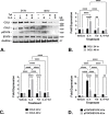
N1 immortalized human prostate fibroblasts were treated with vehicle, IL-4 (20ng/ml), or IL-13 (20 ng/m) with or without 2 hr pre-treatment with IL4Rα (IL4R) (400 ng) or IL-13Rα1 (40 ng) antibodies. Both IL-4 (Fig 7A) and IL-13 (Fig 7D) robustly and significantly induced STAT6 phosphoryation compared to vehicle-treated cells. IL-4 stimulation of STAT6 phosphorylation was repressed upon pre-treatment with IL4Rα (A) but not IL-13Rα1 (B) antibodies, whereas IL-13 stimulation of STAT6 phosphorylation was repressed upon pre-treatment with either IL-13Rα1 (D) or IL4Rα (E) antibodies. Densitometric data (S7 Fig) from the replicate experiments is graphed in C (IL-4) and F (IL-13). Significant differences are indicated as * p < .05; ** p < .01; *** p < .001; **** p < .0001.
Fig 8
Fig 8. IL-4-mediated induction of collagen 1 and collagen 3 expression is coupled to JAK/STAT signaling.
N1 immortalized human prostate fibroblasts were serum-starved for 48 hr then treated with vehicle or IL-4 (20ng/ml) with or without 2 hr pre-treatment with 5um tofacitinib, a JAK2/JAK3 inhibitor, and maintained in serum-free media for an additional 24 or 48 hr. IL-4 significantly induced expression of collagen I (COL1) and collagen 3 (COL3) concurrent with induction of STAT6 phosphorylation, all of which was repressed upon pre-treatment with tofactinib (A). Western blots shown in A are representative of 3 replicate experiments. Densitometric data from the replicate experiments is graphed in B (COL1), C (COL3), and D (pSTAT6/STAT6) (S8 Fig). Significant differences are indicated as * p < .05; ** p < .01; *** p < .001; **** p < .0001.

Similar articles

Cited by

References

    1. Laborde EE, McVary KT. Medical management of lower urinary tract symptoms. Rev Urol. 2009;11(Suppl 1):S19–25. Epub 2010/02/04. ; PubMed Central PMCID: PMC2812890. - PMC - PubMed
    1. Wei JT, Calhoun E, Jacobsen SJ. Urologic diseases in America project: benign prostatic hyperplasia. J Urol. 2005;173(4):1256–61. Epub 2005/03/11. S0022-5347(05)61064-6 [pii] doi: 10.1097/01.ju.0000155709.37840.fe . - DOI - PubMed
    1. Kupelian V, Wei JT, O’Leary MP, Kusek JW, Litman HJ, Link CL, et al.. Prevalence of lower urinary tract symptoms and effect on quality of life in a racially and ethnically diverse random sample: the Boston Area Community Health (BACH) Survey. Arch Intern Med. 2006;166(21):2381–7. Epub 2006/11/30. 166/21/2381 [pii] doi: 10.1001/archinte.166.21.2381 . - DOI - PubMed
    1. Irwin DE, Kopp ZS, Agatep B, Milsom I, Abrams P. Worldwide prevalence estimates of lower urinary tract symptoms, overactive bladder, urinary incontinence and bladder outlet obstruction. BJU International. 2011;108(7):1132–8. doi: 10.1111/j.1464-410X.2010.09993.x - DOI - PubMed
    1. Strope SA, Yang L, Nepple KG, Andriole GL, Owens PL. Population based comparative effectiveness of transurethral resection of the prostate and laser therapy for benign prostatic hyperplasia. J Urol. 2012;187(4):1341–5. doi: 10.1016/j.juro.2011.11.102 ; PubMed Central PMCID: PMC3461307. - DOI - PMC - PubMed

Publication types

MeSH terms