. 2022 Oct 6;17(10):e0274742.
doi: 10.1371/journal.pone.0274742. eCollection 2022.

Therapeutic efficacy of rifalazil (KRM-1648) in a M. ulcerans-induced Buruli ulcer mouse model

Affiliations

Therapeutic efficacy of rifalazil (KRM-1648) in a M. ulcerans-induced Buruli ulcer mouse model

Hanako Fukano et al. PLoS One. .

Abstract

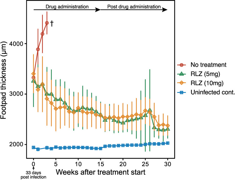
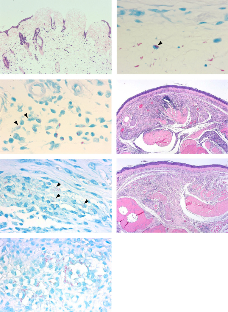
Buruli ulcer (BU) is a skin disease caused by Mycobacterium ulcerans infection that requires long-term antibiotic treatment and/or surgical excision. In this study, we investigated the therapeutic efficacy of the rifamycin derivative, rifalazil (RLZ) (also known as KRM-1648), in an advanced M. ulcerans infection model. Six-week-old female BALB/c mice were infected with 3.25 x 10⁴ colony-forming units (CFU) of M. ulcerans subcutaneously into the bilateral hind footpads. At 33 days post-infection, when the footpads exhibited significant redness and swelling, mice were treated orally with 5 or 10 mg/kg of RLZ for up to 15 weeks. Mice were followed for an additional 15 weeks following treatment cessation. Untreated mice exhibited a progressive increase in footpad redness, swelling, and erosion over time, and all untreated mice reached to endpoint within 5-8 weeks post-bacterial injection. In the RLZ-treated mice, footpad redness and swelling and general condition improved or completely healed, and no recurrence occurred following treatment cessation. After 3 weeks of treatment, the CFU counts from the footpads of recovered RLZ-treated mice showed a 104 decrease compared with those of untreated mice. We observed a further reduction in CFU counts to the detection limit following 6 to 15 weeks of treatment, which did not increase 15 weeks after discontinuing the treatment. Histopathologically, bacteria in the treated mice became fragmented one week after RLZ-treatment. At the final point of the experiment, all the treated mice (5mg/kg/day; n = 6, 10mg/kg/day; n = 7) survived and had no signs of M. ulcerans infection. These results indicate that the rifamycin analogue, RLZ, is efficacious in the treatment of an advanced M. ulcerans infection mouse model.


Conflict of interest statement

No

Figures

Fig 1
Fig 1. Successive measurement of hind footpad thickness.
Measurements were started 33 days post-infection. † All of the mice in the untreated group reached to the endpoint at 4 weeks after the measurement was started.
Fig 2
Fig 2. Log CFU numbers in the hind footpads.
† All of the mice in the untreated group reached to the endpoint at 4 weeks after the measurement was started. N.D Colony was not detected.
Fig 3
Fig 3. Histopathological features of hind footpads.
A-B) Untreated control mouse at day 33 post-infection. A) Erosion, edema, and fibrinous exudates are observed (H&E x290). B) Aggregated acid-fast bacilli are observed primarily in the stroma, with some present in monocytes (arrowhead) (Fite-Faraco Staining x1750). C) Rifalazil (5mg/kg)-treated mouse at one-week of treatment. Some bacterial cells are fragmented (arrowheads) (Fite-Faraco staining x1750). D-E) Rifalazil (5mg/kg)-treated mouse at 15-weeks of treatment. D) Epithelioid cell granuloma with mild infiltration of lymphocytes is noted. Epidermal erosion, edema, fibrin, and neutrophil aggregation are not observed (H&E x120). E) Granularly degenerated acid-fast bacilli are observed in monocytes (arrowheads) (Fite-Faraco staining x1750). F-G) Rifalazil (5mg/kg)-treated mouse 15 weeks following termination of treatment. F) Epithelioid cell granuloma with mild infiltration of lymphocytes is noted. Epidermal erosion, edema, fibrin, and neutrophil aggregation are not observed (H&E x290). G) Completely degenerated acid-fast bacilli are observed in an epithelioid cell granuloma (Fite-Faraco staining x1750).
Fig 4
Fig 4. Workflow of Harmony software image analysis for intracellular M. ulcerans.
THP-1 cell cytoplasm, nuclei, and bacterial immunostaining input image were shown in Fig 4A. Cytoplasm with nucleus were detected by HCS Cell Mask Deep Red and Hoechst 33258 channel respectively (Fig 4B). Bacterial cells were identified as Alexa 488-staining objects within cytoplasm were specifically detected, and calculated the intensity of them (Fig 4C).
Fig 5
Fig 5. Comparative rifamycin efficacy against intracellular M. ulcerans with THP-1 human macrophage infection model.
The sum of fluorescence intensity (Alexa Flour 488) of intracellular bacterium after 72 hours incubation with RFP10μg/mL, RFP10μg/mL+SM10μg/mL, RLZ 10μg/mL and RLZ 10μg/mL+SM10μg/mL. Data are shown as mean ±SD. *p< 0.05 by unpaired t-test.

Similar articles

Cited by

References

    1. Buruli ulcer (Mycobacterium ulcerans infection). [cited 22 Jul 2020]. Available: https://www.who.int/news-room/fact-sheets/detail/buruli-ulcer-(mycobacte...
    1. Loftus MJ, Tay EL, Globan M, Lavender CJ, Crouch SR, Johnson PDR, et al.. Epidemiology of buruli ulcer infections, Victoria, Australia, 2011–2016. Emerg Infect Dis. 2018;24: 1988–1997. doi: 10.3201/eid2411.171593 - DOI - PMC - PubMed
    1. Omansen TF, Erbowor-Becksen A, Yotsu R, Van Der Werf TS, Tiendrebeogo A, Grout L, et al.. Global epidemiology of Buruli ulcer, 2010–2017, and analysis of 2014 WHO programmatic targets. Emerg Infect Dis. 2019;25: 2183–2190. doi: 10.3201/eid2512.190427 - DOI - PMC - PubMed
    1. Nakanaga K, Ogura Y, Toyoda A, Yoshida M, Fukano H, Fujiwara N, et al.. Naturally occurring a loss of a giant plasmid from Mycobacterium ulcerans subsp. shinshuense makes it non-pathogenic. Sci Rep. 2018;8. doi: 10.1038/s41598-018-26425-1 - DOI - PMC - PubMed
    1. Doig KD, Holt KE, Fyfe JAM, Lavender CJ, Eddyani M, Portaels F, et al.. On the origin of Mycobacterium ulcerans, the causative agent of Buruli ulcer. BMC Genomics. 2012;13: 258. doi: 10.1186/1471-2164-13-258 - DOI - PMC - PubMed

Publication types