. 2022 Oct 11;119(41):e2209624119.
doi: 10.1073/pnas.2209624119. Epub 2022 Oct 6.

Sequestration of gut pathobionts in intraluminal casts, a mechanism to avoid dysregulated T cell activation by pathobionts

Affiliations

Sequestration of gut pathobionts in intraluminal casts, a mechanism to avoid dysregulated T cell activation by pathobionts

Martina Sassone-Corsi et al. Proc Natl Acad Sci U S A. .

Abstract

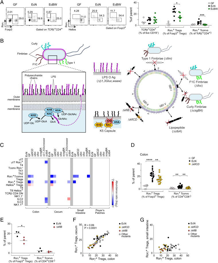
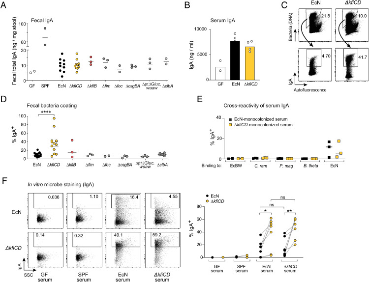
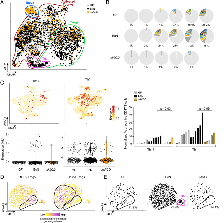
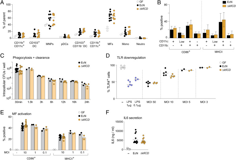
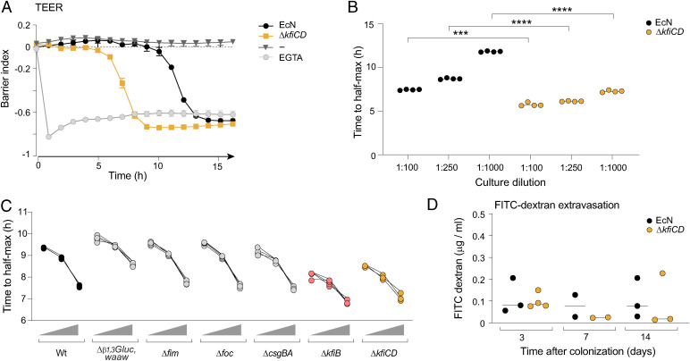
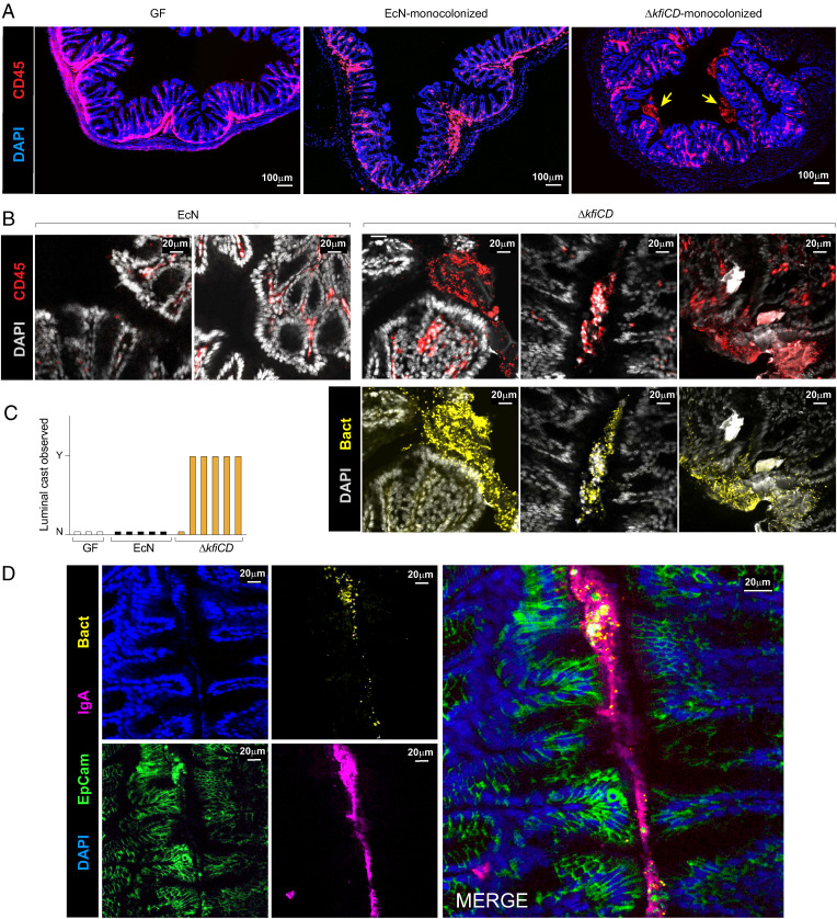
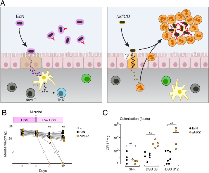
T cells that express the transcription factor RORγ, regulatory (Treg), or conventional (Th17) are strongly influenced by intestinal symbionts. In a genetic approach to identify mechanisms underlying this influence, we performed a screen for microbial genes implicated, in germfree mice monocolonized with Escherichia coli Nissle. The loss of capsule-synthesis genes impaired clonal expansion and differentiation of intestinal RORγ+ T cells. Mechanistic exploration revealed that the capsule-less mutants remained able to induce species-specific immunoglobulin A (IgA) and were highly IgA-coated. They could still trigger myeloid cells, and more effectively damaged epithelial cells in vitro. Unlike wild-type microbes, capsule-less mutants were mostly engulfed in intraluminal casts, large agglomerates composed of myeloid cells extravasated into the gut lumen. We speculate that sequestration in luminal casts of potentially harmful microbes, favored by IgA binding, reduces the immune system's actual exposure, preserving host-microbe equilibrium. The variable immunostimulation by microbes that has been charted in recent years may not solely be conditioned by triggering molecules or metabolites but also by physical limits to immune system exposure.

Keywords: CD4+ T cell; IgA; capsule; genetic screen; gut.


Conflict of interest statement

The authors declare no competing interest.

Figures

Fig. 1.
Fig. 1.
Screening of EcN mutants’ immunomodulatory activities. (A) Representative dot plots and proportions of CD4+ RORγ+ Tconv (Foxp3 RORγ+) and RORγ+ Tregs from colonic LP in germfree and mice monocolonized with EcN and EcBW (4 independent experiments, t test *P value <0.05, ***P value <0.001). (B) Left: schematic illustration of EcN outer membrane structure with capsule polysaccharides (purple) and Kfi components of their biosynthetic pathway. Right: schematic of EcN and EcBW genome comparison (SI Appendix, Fig. S1B), with the positions of the mutants created here and their impact on EcN outer membrane. (C) Average fold changes (at P < 0.01) among immunocytes of different organs of mice monocolonized with the mutants, relative to EcN. (D) Frequencies of RORγ+ Treg and Tconv from colonic LP in GF and mice monocolonized with EcN and capsule mutant ΔkfiCD (five independent experiments, t test **P < 0.01, ****P < 10–4). (E) As (D) for mutant ΔkfiB (*P < 0.05). (F, G) Frequencies of RORγ+ Tregs in colon, cecum, and small intestine LP.
Fig. 2.
Fig. 2.
EcN capsule-less Δkfi mutant is highly coated with specific IgA. (A) Total IgA (from ELISA) in stool of germ-free and monocolonized mice. (B) Total serum IgA (from ELISA) in germ-free and monocolonized mice (day 14). (C) Representative cytometry profiles and quantification (D) of IgA-coated bacteria in EcN or ΔkfiCD monocolonized mice (t test ****P < 0.0001). (E) Binding of serum IgA from EcN or ΔkfiCD monocolonized mice, assessed by flow cytometry, against EcBW, Clostridium ramosum, Peptostreptococcus magnus, and Bacteroides thetaiotaomicron. (F) Representative cytometry plots and quantification of binding to EcN or ΔkfiCD bacteria of serum IgA from EcN or ΔkfiCD monocolonized mice, GF or SPF serum for reference (paired t test *P < 0.05, **P < 0.01).
Fig. 3.
Fig. 3.
T cell activation and clonal expansion are impaired in mice monocolonized with the ΔkfiCD mutant. scRNA-seq and TCR-seq of GF or EcN and ΔkfiCD monocolonized mice (day14). (A) UMAP projection of transcriptomes from CD4+ T cells; cell populations distinguished by expression of signature gene and modules (per S4A). One representative experiment shown. (B) Proportion of αβ TCR clonotypes in individual mice, GF, or monocolonized. Nonexpanded clones in gray, colored clonotypes present in two or more cells (Dataset S3 for details). (C) Projection on a UMAP dimensionality reduction (activated Tconv cells only) of averaged Th1 and Th17 signatures (listed in Materials and Methods), with corresponding violin plots below. Right: quantification of activated T conv cells positive for Th1 and Th17 gene signatures (normalized to total CD4+ T cells). Each bar corresponds to one mouse (Mann-Whitney P value). (D) UMAP projection (Treg cells from all mice in one experiment), color-coded for expression of RORγ+ and Helios+ Treg gene signatures (genes in Materials and Methods and Dataset S4). (E) As (D), but split between hash tagged cells from different mice; proportion of RORγ+ aTregs and the position of Tis Tregs are shown.
Fig. 4.
Fig. 4.
Myeloid cells are similarly activated by both EcN and its capsule-less mutant. (A) Myeloid cell proportions, measured by flow cytometry, in colonic LP of GF, or EcN and ΔkfiCD monocolonized mice (day 14; gating strategy from (SI Appendix, Fig. S1E)). (B) Activation of colonic LP myeloid cells assessed by expression of CD86 and MHCII expression 24 h after monocolonization with EcN or ΔkfiCD. (C) Phagocytosis and clearance by BMDM in culture at different times after infection with live EcN and ΔkfiCD at a multiplicity of infection (MOI) = 10, evaluated as cell-associated cfu. (D) TLR4 surface expression on immortalized BMDM after 20 min exposure to EcN or ΔkfiCD at different MOI (LPS used as a positive control). Different concentration of LPS was used as positive control. (E) BMDM activation 24 h after exposure to live EcN and ΔkfiCD in vitro, assessed as expression of CD86 and MHCII. (F) As (E), IL-6 concentration measured by ELISA from culture supernatants after 24 h.
Fig. 5.
Fig. 5.
Lack of capsule increases damaging effects on epithelial cells in vitro. (A) Caco-2 cells were cultured in CytoView-Z 96 plates, and TEER was recorded over time after challenge (1:1,000) with EcN or ΔkfiCD (EGTA as a positive control). The barrier index was calculated by “axion impedance module” as the ratio between cellular resistance at low frequency (1 Hz) vs. high frequency (41 Hz). (B) As (A), the time of transition (50% drop in barrier index) was measured in cultures supplemented with EcN or ΔkfiCD at different dilutions. (t test, ***P < 0.001, ****P < 10–4); each point a quadruplicate within an experiment, representative of three experiments. (C) As (B), time to transition after exposure to various EcN mutants at three dilutions (1:1,000, 1:5,000, and 1:104). (D) Assessment of in vivo gut permeability as leakage of FITC dextran (4 kDa) administered by gavage and detected in serum 4 h after oral gavage at different times after monocolonization with EcN or ΔkfiCD (each point a different mouse).
Fig. 6.
Fig. 6.
Luminal IgA-immune cell casts engulf the capsule-less EcN mutant. Confocal imaging of colonic tissue was collected for imaging from GF, EcN, or ΔkfiCD monocolonized mice (day 14). (A) Confocal imaging of colonic tissue immunostained for pan-immunocytes (CD45) and cell nuclei (DAPI). Arrows point to intraluminal CD45+ structures. (B) As (A), with counterstaining for bacteria (anti-mCherry). (C) Quantification of intraluminal casts (scored as presence of discrete CD45+ structures, 100 μm or more) in the colon of GF, EcN, and ΔkfiCD monocolonized mice. Each bar represents an individual mouse. (D) As (B), staining for IgA, Epcam (epithelial cells), and bacteria (anti-mCherry).
Fig. 7.
Fig. 7.
Deleterious effects of the capsule-less mutant during DSS-induced colitis. (A) Schematic representation of proposed model, comparing the epithelial-initiated response to EcN or ΔkfiCD that leads either to RORγ+ cell activation or to myeloid cell extravasation. (B, C) Specific pathogen-free animals were fed 2.5% DSS for 6 d, then gavaged with 107 EcN or ΔkfiCD bacteria (DSS dose lowered to 1.5% for the remaining time). Weight was recorded and normalized to their initial weight. (C) As (B), measure of live bacteria (as cfu/mg of feces) at days 8 or 12 (t test, **P < 0.01).

Similar articles

Cited by

References

    1. Chu H., Mazmanian S. K., Innate immune recognition of the microbiota promotes host-microbial symbiosis. Nat. Immunol. 14, 668–675 (2013). - PMC - PubMed
    1. Ivanov I. I., Tuganbaev T., Skelly A. N., Honda K., T cell responses to the microbiota. Annu. Rev. Immunol. 40, 559–587 (2022). - PMC - PubMed
    1. Cress B. F., et al. , Masquerading microbial pathogens: Capsular polysaccharides mimic host-tissue molecules. FEMS Microbiol. Rev. 38, 660–697 (2014). - PMC - PubMed
    1. Sansonetti P. J., Di Santo J. P., Debugging how bacteria manipulate the immune response. Immunity 26, 149–161 (2007). - PubMed
    1. Diacovich L., Gorvel J. P., Bacterial manipulation of innate immunity to promote infection. Nat. Rev. Microbiol. 8, 117–128 (2010). - PubMed

Publication types

Substances