. 2022 Oct 23;163(12):bqac163.
doi: 10.1210/endocr/bqac163.

Dehydroepiandrosterone Supplementation Results in Varying Tissue-specific Levels of Dihydrotestosterone in Male Mice

Affiliations

Dehydroepiandrosterone Supplementation Results in Varying Tissue-specific Levels of Dihydrotestosterone in Male Mice

Hannah Colldén et al. Endocrinology. .

Erratum in

Abstract

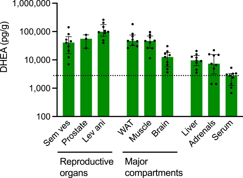
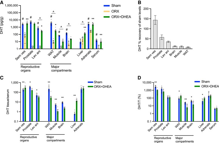
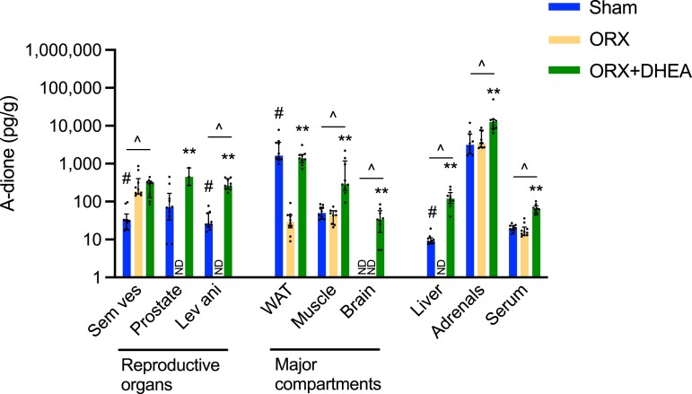
Dehydroepiandrosterone (DHEA), an adrenal androgen precursor, can be metabolized in target tissues into active sex steroids. It has been proposed that DHEA supplementation might result in restoration of physiological local sex steroid levels, but knowledge on the effect of DHEA treatment on local sex steroid levels in multiple tissues is lacking. To determine the effects of DHEA on tissue-specific levels of sex steroids, we treated orchiectomized (ORX) male mice with DHEA for 3 weeks and compared them with vehicle-treated ORX mice and gonadal intact mice. Intra-tissue levels of sex steroids were analyzed in reproductive organs (seminal vesicles, prostate, m. levator ani), major body compartments (white adipose tissue, skeletal muscle, and brain), adrenals, liver, and serum using a sensitive and validated gas chromatography-mass spectrometry method. DHEA treatment restored levels of both testosterone (T) and dihydrotestosterone (DHT) to approximately physiological levels in male reproductive organs. In contrast, this treatment did not increase DHT levels in skeletal muscle or brain. In the liver, DHEA treatment substantially increased levels of T (at least 4-fold) and DHT (+536%, P < 0.01) compared with vehicle-treated ORX mice. In conclusion, we provide a comprehensive map of the effect of DHEA treatment on intra-tissue sex steroid levels in ORX mice with a restoration of physiological levels of androgens in male reproductive organs while DHT levels were not restored in the skeletal muscle or brain. This, and the unexpected supraphysiological androgen levels in the liver, may be a cause for concern considering the uncontrolled use of DHEA.

Keywords: androgens; dehydroepiandrosterone; dihydrotestosterone; intracrinology; mice; reproductive organs.


Figures

Figure 1.
Figure 1.
DHEA levels in tissues and serum as assessed by gas chromatography–tandem mass spectrometry. Young adult orchiectomized male C57BL/6 mice were treated with 1 mg DHEA/day, 5 days a week for 3 weeks, starting 1 week after orchiectomy. Bars and error bars represent median and interquartile range (IQR), the horizontal dotted line represents the median serum level. n = 9-10, for prostate n = 3. Abbreviations: DHEA, dehydroepiandrosterone; lev ani, m. levator ani; sem ves, seminal vesicles; WAT, white adipose tissue.
Figure 2.
Figure 2.
DHT in tissues and serum as assessed by gas chromatography–tandem mass spectrometry. Young adult orchiectomized (ORX) male C57BL/6 mice were treated with 1 mg DHEA/day, 5 days a week for 3 weeks (ORX + DHEA) and compared with sham-operated (Sham) and ORX male mice with vehicle treatment. A, DHT levels in tissues and serum. Bars and error bars represent median and interquartile range (IQR), significant differences according to Kruskal Wallis with Dunn's test are shown as: * = P < 0.05, ** = P < 0.01 ORX + DHEA vs ORX; # = P < 0.05 ORX vs Sham; ^ = P < 0.05 ORX + DHEA vs Sham. B, Recovery of precastration DHT levels in different tissues after treatment with DHEA in ORX male mice. The increase in local DHT levels with DHEA treatment is expressed as a percentage of the average decrease in local DHT levels with ORX. Calculated as (measured ORX + DHEA DHT level − mean ORX DHT level)/(mean Sham DHT level − mean ORX DHT level). Bars and error bars show mean and SEM. C, Ratio of DHT levels in tissue/serum. Significant differences according to Mann-Whitney test are shown as * = P < 0.05, ** P < 0.01. D, Ratio between DHT and T concentrations, an indicator of 5-alpha-reductase activity, in tissues and serum. Significant differences according to Mann-Whitney test are shown as * = P < 0.05, ** P < 0.01. n = 9-10, for prostate n = 3. Data for Sham and ORX control groups were previously published (28). Abbreviations: DHEA, dehydroepiandrosterone; DHT, dihydrotestosterone; lev ani, m. levator ani; sem ves, seminal vesicles; WAT, white adipose tissue; ND, not detected.
Figure 3.
Figure 3.
Testosterone levels in tissues and serum as assessed by gas chromatography–tandem mass spectrometry. Young adult orchiectomized (ORX) male C57BL/6 mice were treated with 1 mg DHEA/day, 5 days a week for 3 weeks (ORX + DHEA) and compared with sham-operated (Sham) and ORX male mice with vehicle treatment. Bars and error bars represent median and interquartile range (IQR), n = 9-10, for prostate n = 3. Significant differences according to Kruskal Wallis with Dunn's test are shown as: * = P < 0.05, ** = P < 0.01 ORX + DHEA vs ORX; # = P < 0.05 ORX vs Sham; ^ = P < 0.05 ORX + DHEA vs Sham. Data for Sham and ORX control groups were previously published (28). Abbreviations: DHEA, dehydroepiandrosterone; sem ves, seminal vesicles; lev ani, m. levator ani; T, testosterone; WAT, white adipose tissue; ND, not detected.
Figure 4.
Figure 4.
Androstenedione levels in tissues and serum as assessed by gas chromatography–tandem mass spectrometry. Young adult orchiectomized (ORX) male C57BL/6 mice were treated with 1 mg DHEA/day, 5 days a week for 3 weeks (ORX + DHEA) and compared with sham-operated (Sham) and ORX male mice with vehicle treatment. Bars and error bars represent median and interquartile range (IQR), n = 9-10, for prostate n = 3. Significant differences according to Kruskal Wallis with Dunn's test are shown as: * = P < 0.05, ** = P < 0.01 ORX + DHEA vs ORX; # = P < 0.05 ORX vs Sham; ^ = P < 0.05 ORX + DHEA vs Sham. Data for Sham and ORX control groups were previously published (28). Abbreviations: A-dione, androstenedione; DHEA, dehydroepiandrosterone; lev ani, m. levator ani; sem ves, seminal vesicles; WAT, white adipose tissue; ND, not detected.

Similar articles

Cited by

References

    1. Li Y, Ren J, Li N, et al. . A dose-response and meta-analysis of dehydroepiandrosterone (DHEA) supplementation on testosterone levels: perinatal prediction of randomized clinical trials. Exp Gerontol. 2020;141:111110. doi:10.1016/j.exger.2020.111110 - DOI - PubMed
    1. Swerdloff RS, Dudley RE, Page ST, Wang C, Salameh WA. Dihydrotestosterone: biochemistry, physiology, and clinical implications of elevated blood levels. Endocr Rev. 2017;38(3):220–254. - PMC - PubMed
    1. Collden H, Nilsson ME, Norlen AK, et al. . Supplemental materials for: Dehydroepiandrosterone supplementation results in varying tissue-specific levels of dihydrotestosterone in male mice. Published September 15, 2022. Zenodo. 10.5281/zenodo.7082262 - DOI - PMC - PubMed
    1. Klinge CM, Clark BJ, Prough RA. Dehydroepiandrosterone research: past, current, and future. Vitam Horm. 2018;108:1–28. doi:10.1016/bs.vh.2018.02.002 - DOI - PubMed
    1. Uhlén M, Fagerberg L, Hallström BM, et al. . Proteomics. Tissue-based map of the human proteome. Science. 2015;347(6220):1260419. - PubMed

Publication types