. 2023 Jan 1;72(1):85-96.
doi: 10.2337/db22-0191.

Recognition of mRNA Splice Variant and Secretory Granule Epitopes by CD4+ T Cells in Type 1 Diabetes

Affiliations

Recognition of mRNA Splice Variant and Secretory Granule Epitopes by CD4+ T Cells in Type 1 Diabetes

Perrin Guyer et al. Diabetes. .

Abstract

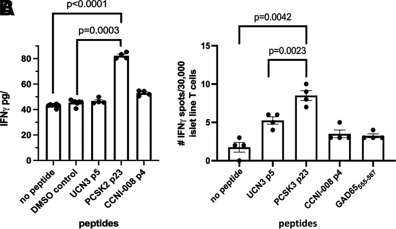
A recent discovery effort resulted in identification of novel splice variant and secretory granule antigens within the HLA class I peptidome of human islets and documentation of their recognition by CD8+ T cells from peripheral blood and human islets. In the current study, we applied a systematic discovery process to identify novel CD4+ T cell epitopes derived from these candidate antigens. We predicted 145 potential epitopes spanning unique splice junctions and within conventional secretory granule antigens and measured their in vitro binding to DRB1*04:01. We generated HLA class II tetramers for the 35 peptides with detectable binding and used these to assess immunogenicity and isolate T cell clones. Tetramers corresponding to peptides with verified immunogenicity were then used to label T cells specific for these putative epitopes in peripheral blood. T cells that recognize distinct epitopes derived from a cyclin I splice variant, neuroendocrine convertase 2, and urocortin-3 were detected at frequencies that were similar to those of an immunodominant proinsulin epitope. Cells specific for these novel epitopes predominantly exhibited a Th1-like surface phenotype. Among the three epitopes, responses to the cyclin I peptide exhibited a distinct memory profile. Responses to neuroendocrine convertase 2 were detected among pancreatic infiltrating T cells. These results further establish the contribution of unconventional antigens to the loss of tolerance in autoimmune diabetes.


Figures

Figure 1
Figure 1
Recognition of novel epitopes by T cells from subjects with type 1 diabetes. A: T cells that recognize epitopes derived from splice variants and novel secretory granule antigens could be expanded from peripheral blood after peptide-specific expansion and detected by HLA class II tetramer staining. Representative results from one individual donor per epitope are shown (out of 15 subjects with T1D, Supplementary Table 1). B: Epitope-specific T cell clones were isolated by sorting and expanding single tetramer–positive T cells. Each clone proliferated robustly (stimulation index >100) in response to 10 µg/mL of its cognate peptide and exhibited negligible proliferation (stimulation index <2) in response to 10 µg/mL of an irrelevant peptide. Data are represented as stimulation index values, which we calculated by normalizing the proliferation of each clone based on [3H]-thymidine incorporation of unstimulated wells. Irrel., Irrelevant; Rel., Relevant; Tmr, Tetramer.
Figure 2
Figure 2
T cells specific for novel epitopes have variable frequencies in subjects with type 1 diabetes. T cells specific for the novel epitopes identified were directly enumerated in peripheral blood using a magnetic enrichment procedure. A: HLA class II tetramers were used to enumerate CD4+ T cells for three novel epitopes plus a reference PPI epitope. Cells were gated based on size, viability, and lack of CD14/CD19 expression and CD4 expression and then displayed on a CD4 vs. tetramer (PE, PE-CF594, PE-Cy5, or PE-Cy7 labeled). A typical result, representative of 10 subjects with T1D examined (Supplementary Table 2), is shown. Each upper panel shows a precolumn fraction, used to determine the total number of CD4+ T cells in the unmanipulated sample and to set a threshold for positive tetramer staining. Each lower panel shows the corresponding enriched (postcolumn) fraction, used to determine the total number of epitope-specific CD4+ T cells in the sample. B: Summary of ex vivo frequencies for each epitope measured in the peripheral blood of 10 subjects with established type 1 diabetes (Supplementary Table 2). The dotted line indicates the previously reported limit of detection for direct tetramer staining. CCNI and PPI were more frequent (5.6 and 5.3 cells per million, respectively) than PCSK2 and UCN3 (3.9 and 3.6 cells per million). C: A heat map of the same frequency data reveals unique patterns of reactivity in different subjects with type 1 diabetes. Post, enriched fractions; Pre, nonenriched fractions; Tmr, Tetramer.
Figure 3
Figure 3
T cells specific for novel epitopes exhibit different phenotypes. Epitope-specific CD4+ T cells in subjects with T1D (Supplementary Table 2) were phenotyped based on their cell surface expression of lineage markers. A: CD45RA and CCR7 surface staining was used to classify T cells as naive (CD45RA+CCR7+), central memory (CD45RACCR7+), effector memory (CD45RACCR7), or terminal effectors (CD45RA+CCR7). This allowed comparison of the memory distributions for CCNI, UCN3, PCSK2, and PPI (as a reference epitope). B: Chemokine receptors were used to subdivide memory T cells into Th1-like (CXCR3+, CCR4, and CCR6), Th2-like (CXCR3, CCR4+, and CCR6), Th17-like (CXCR3+, CCR4, and CCR6+), Th1/17-like (CXCR3+, CCR4, and CCR6+), Th1/2-like (CXCR3+, CCR4+, and CCR6), or Th1*-like (CXCR3+, CCR4+, and CCR6+) states. This allowed comparison of the cell subset distributions for CCNI, UCN3, PCSK2, and PPI (as a reference epitope). C: CCNI had a distinct memory profile, with a significantly lower proportion of naive cells than UCN3 (P = 0.04) and trended lower than PCSK2 (P = 0.07). UCN3 had a distinct cell subset distribution, with a significantly lower proportion of Th1-like cells than CCNI (P = 0.04) and proportion of Th2-like cells that was significantly higher than PCSK2 (P = 0.03) and trended higher than CCNI (P = 0.1). CM, central memory; EM, effector memory.
Figure 4
Figure 4
T cells specific for novel epitopes are more frequent in subjects with type 1 diabetes and exhibit phenotype differences. HLA class II tetramers were used to characterize T cells specific for the three novel epitopes in 10 HLA-matched control subjects (Supplementary Table 3). A: Combining all three epitopes (CCNI, PCSK2, and UCN3) total frequencies were significantly higher (P = 0.0003, Mann Whitney test) in subjects with type 1 diabetes (black circles) than in HLA-matched controls (white circles). B. In examining individual epitopes, individual frequencies were significantly higher in subjects with type 1 diabetes (black circles, taken from Fig. 2B but shown here on a log axis to more effectively depict the low frequencies seen in control subjects) than in healthy control subjects (white circles) only for CCNI (P = 0.021). C: With combination of all three epitopes (CCNI, PCSK2, and UCN3) the proportion of T cells that had a memory phenotype (calculated as a ratio of the combined percentage of all memory subsets and percentage that were naive) was not significantly different (P = 0.565, Mann Whitney U test) in subjects with type 1 diabetes (black circles) than in HLA-matched control subjects (white circles). D: Chemokine receptors were used to subdivide memory T cells from healthy control subjects into Th1-like, Th2-like, Th17-like, Th1/17-like, Th1/2-like, or Th1*-like states (just as in Fig. 3B for subjects with T1D). Control subjects lacked the Th1-like bias seen for some specificities in subjects with type 1 diabetes.
Figure 5
Figure 5
PCSK2-specific CD4+ T cells infiltrate the islets of a donor with type 1 diabetes. An islet-derived CD4+ T cell line for a donor with T1D recognizes PCSK2. T cells were isolated and expanded from live, vibratome slices of pancreas tissue from nPOD donor 6472. A: Irradiated autologous B-LCL pulsed with peptides at 50 µg/mL, no peptide, or DMSO volume control and then cultured with T cells and anti-CD28 (5 µg/mL) for costimulation for 48 h. Supernatants were collected and responses detected with a standard IFN-γ ELISA. The data shown represent one of four similar experiments. B: The islet T cell line previously cultured with PCSK3 peptide was restimulated (with peptide-pulsed autologous B-LCL as antigen-presenting cells), expanded, and then retested by IFNγ ELISpot for reactivity to the peptides. Tumor necrosis factor-α was not detected by ELISpot. The data shown represent one of two similar experiments.

Similar articles

Cited by

References

    1. Coppieters KT, Dotta F, Amirian N, et al. . Demonstration of islet-autoreactive CD8 T cells in insulitic lesions from recent onset and long-term type 1 diabetes patients. J Exp Med 2012;209:51–60 - PMC - PubMed
    1. Di Lorenzo TP, Peakman M, Roep BO. Translational mini-review series on type 1 diabetes: Systematic analysis of T cell epitopes in autoimmune diabetes. Clin Exp Immunol 2007;148:1–16 - PMC - PubMed
    1. Roep BO, Peakman M. Antigen targets of type 1 diabetes autoimmunity. Cold Spring Harb Perspect Med 2012;2:a007781. - PMC - PubMed
    1. Erlich H, Valdes AM, Noble J, et al. .; Type 1 Diabetes Genetics Consortium . HLA DR-DQ haplotypes and genotypes and type 1 diabetes risk: analysis of the type 1 diabetes genetics consortium families. Diabetes 2008;57:1084–1092 - PMC - PubMed
    1. James EA, Mallone R, Kent SC, DiLorenzo TP. T-cell epitopes and neo-epitopes in type 1 diabetes: A comprehensive update and reappraisal. Diabetes 2020;69:1311–1335 - PMC - PubMed

Publication types