. 2022 Nov;298(11):102560.
doi: 10.1016/j.jbc.2022.102560. Epub 2022 Oct 4.

Reconstitution of the SARS-CoV-2 ribonucleosome provides insights into genomic RNA packaging and regulation by phosphorylation

Affiliations

Reconstitution of the SARS-CoV-2 ribonucleosome provides insights into genomic RNA packaging and regulation by phosphorylation

Christopher R Carlson et al. J Biol Chem. 2022 Nov.

Abstract

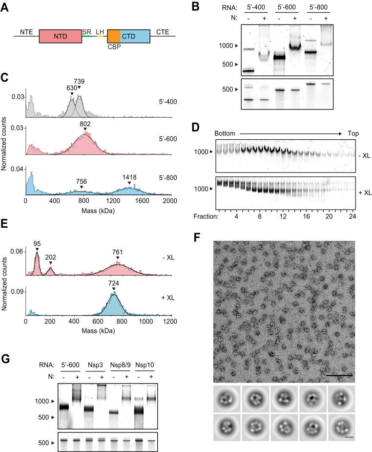
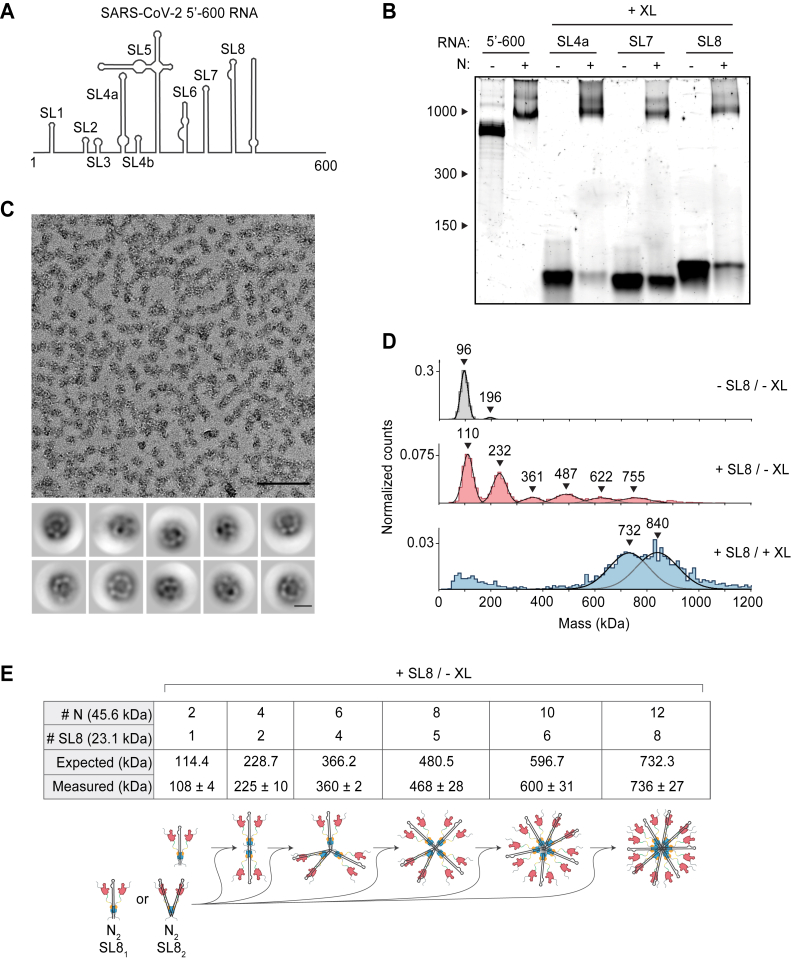
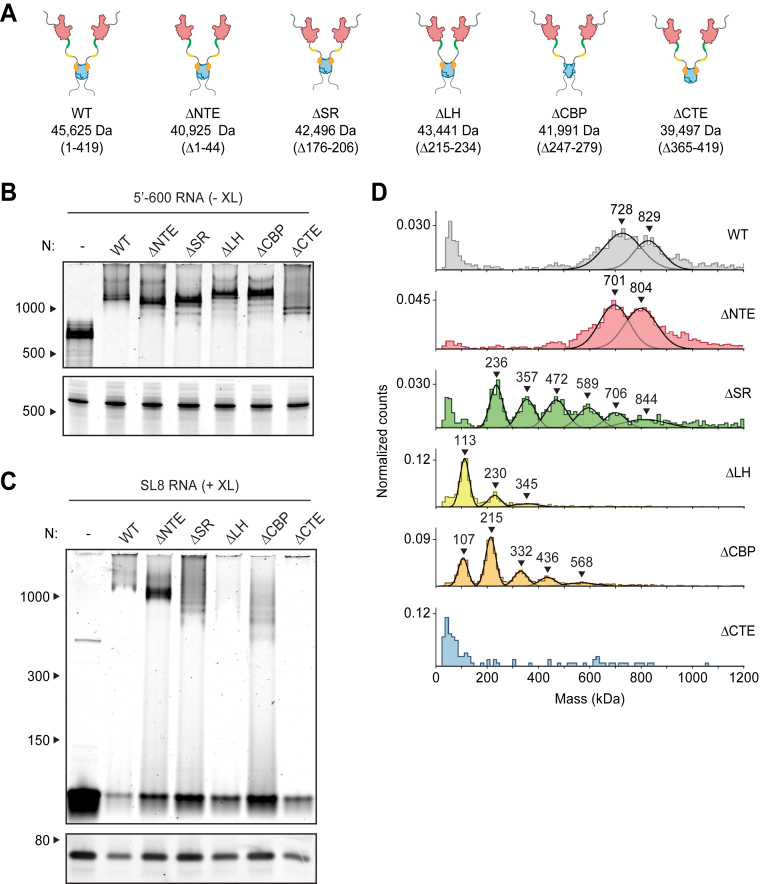
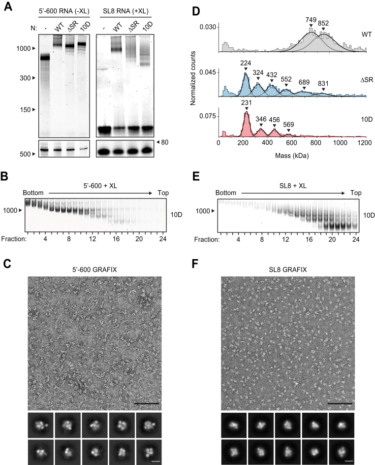
The nucleocapsid (N) protein of severe acute respiratory syndrome coronavirus 2 is responsible for compaction of the ∼30-kb RNA genome in the ∼90-nm virion. Previous studies suggest that each virion contains 35 to 40 viral ribonucleoprotein (vRNP) complexes, or ribonucleosomes, arrayed along the genome. There is, however, little mechanistic understanding of the vRNP complex. Here, we show that N protein, when combined in vitro with short fragments of the viral genome, forms 15-nm particles similar to the vRNP structures observed within virions. These vRNPs depend on regions of N protein that promote protein-RNA and protein-protein interactions. Phosphorylation of N protein in its disordered serine/arginine region weakens these interactions to generate less compact vRNPs. We propose that unmodified N protein binds structurally diverse regions in genomic RNA to form compact vRNPs within the nucleocapsid, while phosphorylation alters vRNP structure to support other N protein functions in viral transcription.

Keywords: N protein; RNA binding protein; RNA virus; SARS-CoV-2; nucleocapsid; nucleosome; phosphorylation; plus-stranded RNA virus.


Conflict of interest statement

Conflict of interest The authors declare that they have no conflicts of interest with the contents of this article.

Figures

Figure 1
Figure 1
Viral RNA promotes formation of the SARS-CoV-2 ribonucleosome.A, schematic of N protein domain architecture, including the N-terminal extension (NTE), N-terminal domain (NTD), serine/arginine region (SR), leucine helix (LH), C-terminal basic patch (CBP), C-terminal domain (CTD), and C-terminal extension (CTE). B, native (top) and denaturing (bottom) PAGE analysis of 15 μM N protein mixed with 256 ng/μl of the indicated RNA, stained with SYBR Gold to detect RNA species. RNA length standards shown on left (nt). RNA concentration in these and other experiments was 256 ng/μl, regardless of RNA length, to ensure that all mixtures contain the same nucleotide concentration. C, mass photometry analysis of vRNP complexes formed in the presence of 15 μM N and 256 ng/μl RNA. Data were fit to Gaussian distributions, with mean molecular mass indicated above each peak. Representative of two independent experiments (Table S1). D, native gel analysis of glycerol gradient separated vRNP complexes. Top, no crosslinker added (−XL); bottom: 0.1% glutaraldehyde added (+XL) to 40% glycerol buffer (GraFix). RNA length standard shown on left (nt). E, fractions 7 and 8 (from D) were combined and analyzed by mass photometry, as in C. Top, no crosslinker (−XL); bottom, GraFix-purified vRNP (+XL). Representative of two independent experiments (Table S1). F, negative stain electron microscopy and two-dimensional classification of GraFix-purified vRNPs (combined fractions 7 and 8 from D). Scale bars are 100 nm (top) and 10 nm (bottom). G, native (top) and denaturing gel analysis (bottom) of 15 μM N protein mixed with 256 ng/μl of the indicated 600 nt RNA molecules. RNA length standards shown on left (nt). See Table S2 for sequences. N, nucleocapsid; vRNP, viral ribonucleoprotein.
Figure 2
Figure 2
Stem-loop RNA, in complex with N protein, drives ribonucleosome formation.A, schematic of RNA secondary structure in the 5′-600 RNA (46). B, native gel analysis of 15 μM N protein mixed with 256 ng/μl of the indicated RNAs. Samples containing stem-loop RNA were crosslinked to stabilize the resulting complex, while the 5′-600 RNA sample was left un-crosslinked. RNA length standards shown on left (nt). Corresponding denaturing gel analysis shown in Fig. S1A. C, fractions 7 and 8 of GraFix-purified SL8 assembled vRNPs were combined and analyzed by negative stain electron microscopy and two-dimensional classification. Scale bars are 100 nm (top) and 10 nm (bottom). D, mass photometry analysis of indicated N protein-RNA mixtures. Top, N protein alone; middle, N protein mixed with SL8, un-crosslinked; bottom, crosslinked complexes of N protein bound to SL8 (data reproduced from Fig. S1B for ease of comparison). Representative of two independent experiments (Table S1). E, predictions of N protein and RNA stoichiometry, based on measured masses of N protein in complex with SL8 RNA without crosslinker (D, middle panel). Measured masses are means ± standard deviation in two independent experiments (Table S1). Below the table is a schematic of a proposed assembly mechanism in which N protein dimers, bound to one or two stem-loop RNAs, iteratively assemble to the full vRNP. N, nucleocapsid; vRNP, viral ribonucleoprotein.
Figure 3
Figure 3
Multiple N protein regions contribute to vRNP formation.A, schematic of wildtype (WT) N protein and deletion mutants, as described in the text. Mass is that of monomeric N protein. B, 15 μM N protein mutants were mixed with 256 ng/μl 5′-600 RNA and analyzed by native (top) and denaturing (bottom) gel electrophoresis. RNA length standards shown on left (nt). C, 20 μM N protein mutants were mixed with 256 ng/μl SL8 RNA, crosslinked, and analyzed by native (top) and denaturing (bottom) gel electrophoresis. RNA length standards shown on left (nt). D, mass photometry analysis of crosslinked N protein mutants (20 μM) bound to SL8 RNA (256 ng/μl). Representative of at least two independent experiments (Table S1). CBP, C-terminal basic patch; CTD, C-terminal domain; CTE, C-terminal extension; LH, leucine helix; N, nucleocapsid; NTD, N-terminal domain; NTE, N-terminal extension; SR, serine/arginine region; vRNP, viral ribonucleoprotein.
Figure 4
Figure 4
Phosphomimetic mutations in the SR region of N prevent vRNP assembly.A, 15 μM N protein constructs were combined with 256 ng/μl 5′-600 RNA (left) or SL8 RNA (right) and analyzed by native (top) and denaturing (bottom) gel electrophoresis. SL8 ribonucleoprotein complexes were crosslinked prior to native gel electrophoresis. RNA length standards shown on left (nt). See Figure 5A for the ten sites of phosphorylation mutated to aspartic acid in the 10D mutant. B, 15 μM phosphomimetic N protein (10D) was mixed with 256 ng/μl 5′-600 RNA and separated by glycerol gradient centrifugation in the presence of crosslinker (GraFix). Fractions were collected and analyzed by native gel electrophoresis. RNA length standard shown on left (nt). C, fractions 7 and 8 of GraFix-separated vRNPs (from B) were combined and analyzed by negative stain electron microscopy and two-dimensional classification. Scale bars are 100 nm (top) and 10 nm (bottom). D, 15 μM N protein mutants were mixed with 256 ng/μl SL8 RNA, crosslinked, and analyzed by mass photometry. Representative of at least two independent experiments (Table S1). A separate analysis of ΔSR mutant is also shown in Figure 3D. E, 15 μM 10D N protein was mixed with 256 ng/μl SL8 RNA and separated by GraFix. Fractions were analyzed by native gel electrophoresis. F, fractions 19 and 20 of GraFix-purified 10D N in complex with SL8 RNA (from E) were combined and visualized by negative stain electron microscopy and two-dimensional classification. Scale bars are 100 nm (top) and 10 nm (bottom). N, nucleocapsid; SR, serine/arginine region; vRNP, viral ribonucleoprotein; WT, wild type.
Figure 5
Figure 5
Phosphorylation of N protein inhibits ribonucleosome formation.A, sequence of N protein SR regions from SARS-CoV (aa 177–210) and SARS-CoV-2 (aa 176–209). The proposed mechanism of sequential phosphorylation (30) is initiated by SRPK at S188 and S206 (orange), which leads to upstream phosphorylation of eight sites by GSK3 (green), allowing for final phosphorylation of four additional sites by CK1 (purple). In the phosphomimetic 10D mutant used in Figure 4, the SRPK and GSK3 sites are changed to aspartic acid. B, wildtype (WT) and mutant N protein constructs were incubated with the indicated kinases in the presence of radiolabeled ATP and analyzed by SDS-PAGE and autoradiography. Phosphorylated N is indicated. Asterisk denotes autophosphorylation of CK1. Molecular mass marker shown on right (kDa). C, N protein (WT or S188A + S206A) was phosphorylated by SRPK, GSK3, and CK1 and then mixed with SL8 RNA. The resulting ribonucleoprotein complexes were separated by glycerol gradient centrifugation in the presence of crosslinker (GraFix) and analyzed by native gel electrophoresis. RNA length standard shown on left (nt). D, peak fractions from the GraFix analyses in C were analyzed by mass photometry. Top, fractions 19 + 20 of wildtype N; bottom, fractions 7 + 8 of S188A + S206A mutant N. Representative of two independent experiments (Table S1). CK1, casein kinase 1; GSK3, glycogen-synthase kinase 3; N, nucleocapsid; SR, serine/arginine region; SRPK, serine-arginine protein kinase.

Update of

Similar articles

Cited by

References

    1. Zhou P., Yang X.L., Wang X.G., Hu B., Zhang L., Zhang W., et al. A pneumonia outbreak associated with a new coronavirus of probable bat origin. Nature. 2020;579:270–273. - PMC - PubMed
    1. Fung T.S., Liu D.X. Human coronavirus: host-pathogen interaction. Annu. Rev. Microbiol. 2019;73:529–557. - PubMed
    1. Masters P.S. The molecular biology of coronaviruses. Adv. Virus Res. 2006;66:193–292. - PMC - PubMed
    1. Scherer K.M., Mascheroni L., Carnell G.W., Wunderlich L.C.S., Makarchuk S., Brockhoff M., et al. SARS-CoV-2 nucleocapsid protein adheres to replication organelles before viral assembly at the Golgi/ERGIC and lysosome-mediated egress. Sci. Adv. 2022;8:eabl4895. - PMC - PubMed
    1. V'Kovski P., Kratzel A., Steiner S., Stalder H., Thiel V. Coronavirus biology and replication: implications for SARS-CoV-2. Nat. Rev. Microbiol. 2021;19:155–170. - PMC - PubMed

Publication types