

Association between attention-deficit/hyperactivity disorder symptom severity and white matter integrity moderated by in-scanner head motion
- PMID: 36202807
- PMCID: PMC9537185
- DOI: 10.1038/s41398-022-02117-3
Association between attention-deficit/hyperactivity disorder symptom severity and white matter integrity moderated by in-scanner head motion
Abstract
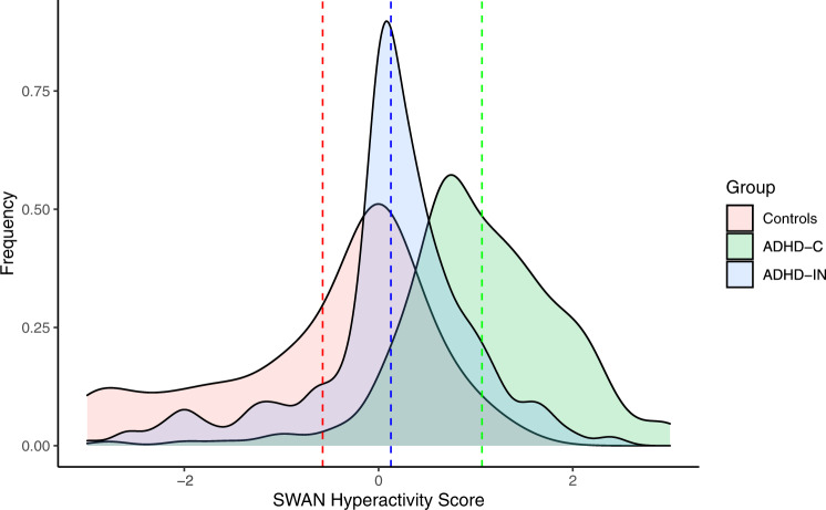
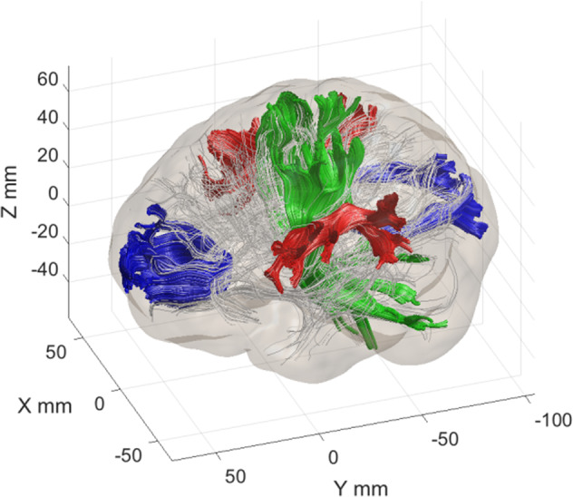
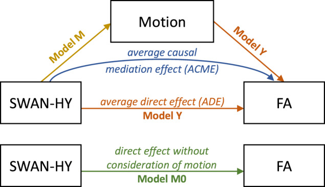
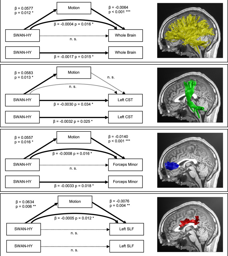
Attention-deficit/hyperactivity disorder (ADHD) is a common and debilitating neurodevelopmental disorder associated with various negative life impacts. The manifestation of ADHD is very heterogeneous, and previous investigations on neuroanatomical alterations in ADHD have yielded inconsistent results. We investigated the mediating effect of in-scanner head motion and ADHD hyperactivity severity on motion-corrected fractional anisotropy (FA) using diffusion tensor imaging in the currently largest sample (n = 739) of medication-naïve children and adolescents (age range 5-22 years). We used automated tractography to examine whole-brain and mean FA of the tracts most frequently reported in ADHD; corpus callosum forceps major and forceps minor, left and right superior-longitudinal fasciculus, and left and right corticospinal tract (CST). Associations between FA and hyperactivity severity appeared when in-scanner head motion was not accounted for as mediator. However, causal mediation analysis revealed that these effects are fully mediated through in-scanner head motion for whole-brain FA, the corpus callosum forceps minor, and left superior-longitudinal fasciculus. Direct effect of hyperactivity severity on FA was only found for the left CST. This study illustrates the crucial role of in-scanner head motion in the identification of white matter integrity alterations in ADHD and shows how neglecting irremediable motion artifacts causes spurious findings. When the mediating effect of in-scanner head motion on FA is accounted for, an association between hyperactivity severity and FA is only present for the left CST; this may play a crucial role in the manifestation of hyperactivity and impulsivity symptoms in ADHD.
© 2022. The Author(s).
Conflict of interest statement
The authors declare no competing interests.
Figures
Similar articles
-
Aberrant white matter properties of the callosal tracts implicated in girls with attention-deficit/hyperactivity disorder.Brain Imaging Behav. 2020 Jun;14(3):728-735. doi: 10.1007/s11682-018-0010-2. Brain Imaging Behav. 2020. PMID: 30556106
-
Association of White Matter Structure With Autism Spectrum Disorder and Attention-Deficit/Hyperactivity Disorder.JAMA Psychiatry. 2017 Nov 1;74(11):1120-1128. doi: 10.1001/jamapsychiatry.2017.2573. JAMA Psychiatry. 2017. PMID: 28877317 Free PMC article.
-
White matter endophenotype candidates for ADHD: a diffusion imaging tractography study with sibling design.Psychol Med. 2020 May;50(7):1203-1213. doi: 10.1017/S0033291719001120. Epub 2019 May 22. Psychol Med. 2020. PMID: 31115278 Clinical Trial.
-
Research Review: Diffusion tensor imaging studies of attention-deficit/hyperactivity disorder: meta-analyses and reflections on head motion.J Child Psychol Psychiatry. 2018 Mar;59(3):193-202. doi: 10.1111/jcpp.12778. Epub 2017 Jul 3. J Child Psychol Psychiatry. 2018. PMID: 28671333
-
White matter alterations in Attention-Deficit/Hyperactivity Disorder (ADHD): a systematic review of 129 diffusion imaging studies with meta-analysis.Mol Psychiatry. 2023 Oct;28(10):4098-4123. doi: 10.1038/s41380-023-02173-1. Epub 2023 Jul 21. Mol Psychiatry. 2023. PMID: 37479785 Free PMC article.
Cited by
-
White matter microstructure in school-age children with down syndrome.Dev Cogn Neurosci. 2025 Jun;73:101540. doi: 10.1016/j.dcn.2025.101540. Epub 2025 Mar 1. Dev Cogn Neurosci. 2025. PMID: 40043413 Free PMC article.
-
Changes in MRI head motion across development: typical development and ADHD.Brain Imaging Behav. 2024 Oct;18(5):1144-1152. doi: 10.1007/s11682-024-00910-w. Epub 2024 Aug 27. Brain Imaging Behav. 2024. PMID: 39190098 Free PMC article.
References
-
- American Psychiatric Association. Diagnostic and Statistical Manual of Mental Disorders (DSM-5®). American Psychiatric Pub, 2013.
MeSH terms
Grants and funding
LinkOut - more resources
Full Text Sources
Medical
