Review
. 2022 Oct 6;8(1):42.
doi: 10.1038/s41526-022-00228-1.

Optic nerve sheath diameter and spaceflight: defining shortcomings and future directions

Affiliations
Review

Optic nerve sheath diameter and spaceflight: defining shortcomings and future directions

Dylan A Fall et al. NPJ Microgravity. .

Abstract

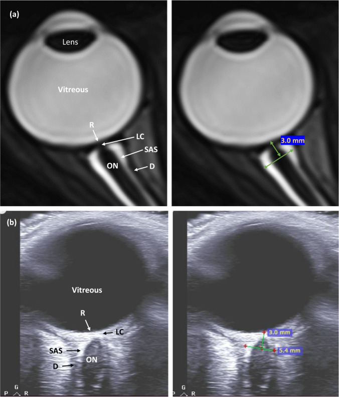
Neuro-ocular changes during long-duration space flight are known as spaceflight-associated neuro-ocular syndrome (SANS). The ability to detect, monitor, and prevent SANS is a priority of current space medicine research efforts. Optic nerve sheath diameter (ONSD) measurement has been used both terrestrially and in microgravity as a proxy for measurements of elevated intracranial pressure. ONSD shows promise as a potential method of identifying and quantitating neuro-ocular changes during space flight. This review examines 13 studies measuring ONSD and its relationship to microgravity exposure or ground-based analogs, including head-down tilt, dry immersion, or animal models. The goal of this correspondence is to describe heterogeneity in the use of ONSD in the current SANS literature and make recommendations to reduce heterogeneity in future studies through standardization of imaging modalities, measurement techniques, and other aspects of study design.


Conflict of interest statement

The authors declare no competing interests.

Figures

Fig. 1
Fig. 1. Example of anatomy and correct measurement of ONSD.
Both MRI (a) and US (b) are represented in the figure. Note the measurement was conducted on a plane where the hypoechoic arachnoid and dural layer are clearly distinguished from the hyperechoic retroorbital face and subarachnoid space. R Retina, LC lamina cribrosa, ON optic nerve, SAS subarachnoid space, D dura.

References

    1. Mader TH, et al. Optic disc edema, globe flattening, choroidal folds, and hyperopic shifts observed in astronauts after long-duration space flight. Ophthalmology. 2011;118:2058–2069. doi: 10.1016/j.ophtha.2011.06.021. - DOI - PubMed
    1. Lee, A. G. et al. Spaceflight associated neuro-ocular syndrome (SANS) and the neuro-ophthalmologic effects of microgravity: a review and an update. npj Microgravity6. 10.1038/s41526-020-0097-9 (2020). - PMC - PubMed
    1. Lee AG, Mader TH, Gibson CR, Brunstetter TJ, Tarver WJ. Space flight-associated neuro-ocular syndrome (SANS) Eye. 2018;32:1164–1167. doi: 10.1038/s41433-018-0070-y. - DOI - PMC - PubMed
    1. Bhargava V, et al. Ultrasonographic optic nerve sheath diameter measurement to detect intracranial hypertension in children with neurological injury: a systematic review. Pediatr. Crit. Care Med. 2020;21:e858–e868. doi: 10.1097/PCC.0000000000002453. - DOI - PubMed
    1. Dubourg J, Javouhey E, Geeraerts T, Messerer M, Kassai B. Ultrasonography of optic nerve sheath diameter for detection of raised intracranial pressure: a systematic review and meta-analysis. Intensiv. Care Med. 2011;37:1059–1068. doi: 10.1007/s00134-011-2224-2. - DOI - PubMed