. 2022 Oct 6;12(1):16189.
doi: 10.1038/s41598-022-19906-x.

Activating effective functional hand movements in individuals with complete tetraplegia through neural stimulation

Affiliations

Activating effective functional hand movements in individuals with complete tetraplegia through neural stimulation

Christine Azevedo Coste et al. Sci Rep. .

Abstract

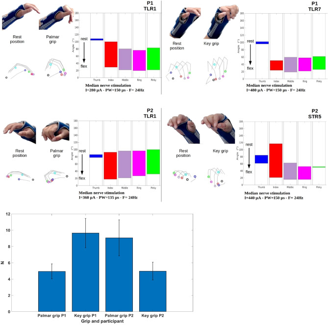
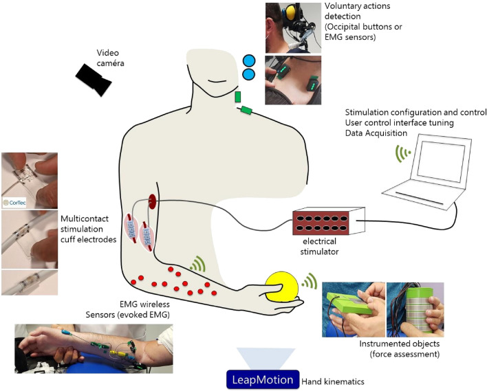
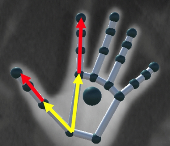
Individuals with complete cervical spinal cord injury suffer from a permanent paralysis of upper limbs which prevents them from achieving most of the activities of daily living. We developed a neuroprosthetic solution to restore hand motor function. Electrical stimulation of the radial and median nerves by means of two epineural electrodes enabled functional movements of paralyzed hands. We demonstrated in two participants with complete tetraplegia that selective stimulation of nerve fascicles by means of optimized spreading of the current over the active contacts of the multicontact epineural electrodes induced functional and powerful grasping movements which remained stable over the 28 days of implantation. We also showed that participants were able to trigger the activation of movements of their paralyzed limb using an intuitive interface controlled by voluntary actions and that they were able to perform useful functional movements such as holding a can and drinking through a straw.

Trial registration: ClinicalTrials.gov NCT04306328.


Conflict of interest statement

David Guiraud and David Andreu are shareholders of NEURINNOV company. There is no other conflict of interest.

Figures

Figure 1
Figure 1
IRO of the muscles for the 3 configurations (TLR, STR, TTR) and for 2 recruitment levels (0.1 and 0.7). From top to bottom: P1, P2 median nerve—P1, P2 radial nerve—Imin values. Each vertex of one colored polygon corresponds to the IRO values for the selected cathode of the considered muscle. The smaller the polygon surface is, the higher the current needed to obtain a recruitment level of 0.1 resp. 0.7 is. A vertex on the circle edge means that Imin, the minimum current amplitude for this configuration, is needed to obtain the given level of recruitment whereas a vertex tied to the center means that the level of recruitment cannot be obtained. In between, along a radius, the sequence of activation with increasing intensities for a given level of recruitment can be seen from edge to center.
Figure 1
Figure 1
IRO of the muscles for the 3 configurations (TLR, STR, TTR) and for 2 recruitment levels (0.1 and 0.7). From top to bottom: P1, P2 median nerve—P1, P2 radial nerve—Imin values. Each vertex of one colored polygon corresponds to the IRO values for the selected cathode of the considered muscle. The smaller the polygon surface is, the higher the current needed to obtain a recruitment level of 0.1 resp. 0.7 is. A vertex on the circle edge means that Imin, the minimum current amplitude for this configuration, is needed to obtain the given level of recruitment whereas a vertex tied to the center means that the level of recruitment cannot be obtained. In between, along a radius, the sequence of activation with increasing intensities for a given level of recruitment can be seen from edge to center.
Figure 2
Figure 2
Recruitment curves of the 3 selected configurations evoking functional movements: Participant P1: TLR2 for hand opening (radial nerve), TLR1 for palmar grip, TLR7 for key grip (median nerve). Participant P2: STR2 for hand opening (radial nerve), TLR1 for palmar grip, STR5 for key grip (median nerve). The green areas show the ranges of usable intensity settings that allow modulating the force while keeping a similar muscles’ synergy.
Figure 3
Figure 3
Kinematics data of hand opening. Left: participant P1—Radial nerve stimulation TLR2 (Wrist brace + Thumb splint). Right: participant P2—Radial nerve stimulation STR2 (Thumb splint). Video snapshots and posture reconstruction based on Leap Motion data. The diagrams represent the excursions of the 5 angles.
Figure 4
Figure 4
Kinematics data of palmar grip with thumb and key grip. Top: Participant P1 (Left: Configuration TLR1, Right: Configuration TLR7). Middle: Participant P2 (Left: Configuration TLR1, Right: Configuration STR5). Video snapshots and posture reconstruction based on Leap Motion data. The diagrams represent the excursions of the 5 angles. Bottom: Normal forces recorded for Palmar (instrumented can) and Key grip (instrumented tablet) for P1 and P2, 3 trials per condition.
Figure 5
Figure 5
Video snapshots illustrating different grasping performances. Left: participant P1. Right: participant P2.
Figure 6
Figure 6
Setup description. An experimental platform was developed to control the stimulation delivered to 2 neural epineural electrodes implanted around the median and radial nerves. Evoked electromyography, video, evoked movement kinematics and grasping forces were recorded. The participants used voluntary muscle contractions or occipital buttons to trigger different stimulation configurations.
Figure 7
Figure 7
For each finger, the angle between the metacarpal segment (yellow arrow) and the extremity of the last phalanx is computed (red arrow). For the thumb the first phalanx is taken into consideration instead of the metacarpal segment.

References

    1. Fitzharris M, Cripps R, Lee B. Estimating the global incidence of traumatic spinal cord injury. Spinal Cord. 2014;52:117. doi: 10.1038/sc.2013.135. - DOI - PubMed
    1. Anderson KD. Targeting recovery: Priorities of the spinal cord-injured population. J. Neurotrauma. 2004;21:1371–1383. doi: 10.1089/neu.2004.21.1371. - DOI - PubMed
    1. Simpson LA, Eng JJ, Hsieh JTC, Wolfe DL, Wolfe and the Spinal Cord Injury Re The health and life priorities of individuals with spinal cord injury: A systematic review. J. Neurotrauma. 2012;29:1548–1555. doi: 10.1089/neu.2011.2226. - DOI - PMC - PubMed
    1. Estores IM. The consumer’s perspective and the professional literature: What do persons with spinal cord injury want? J. Rehabil. Res. Dev. 2003;40:93–98. doi: 10.1682/JRRD.2003.08.0093. - DOI - PubMed
    1. Hanson R, Franklin M. Sexual loss in relation to other functional losses for spinal cord injured males. Arch. Phys. Med. Rehabil. 1976;57:291. - PubMed

Publication types

Associated data