Review
. 2023 Jan;22(1):38-58.
doi: 10.1038/s41573-022-00558-5. Epub 2022 Oct 6.

Targeting replication stress in cancer therapy

Affiliations
Review

Targeting replication stress in cancer therapy

Alexandre André B A da Costa et al. Nat Rev Drug Discov. 2023 Jan.

Abstract

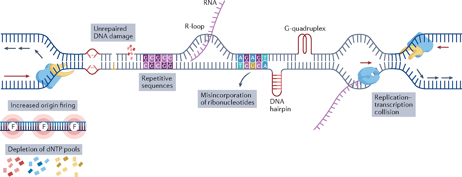
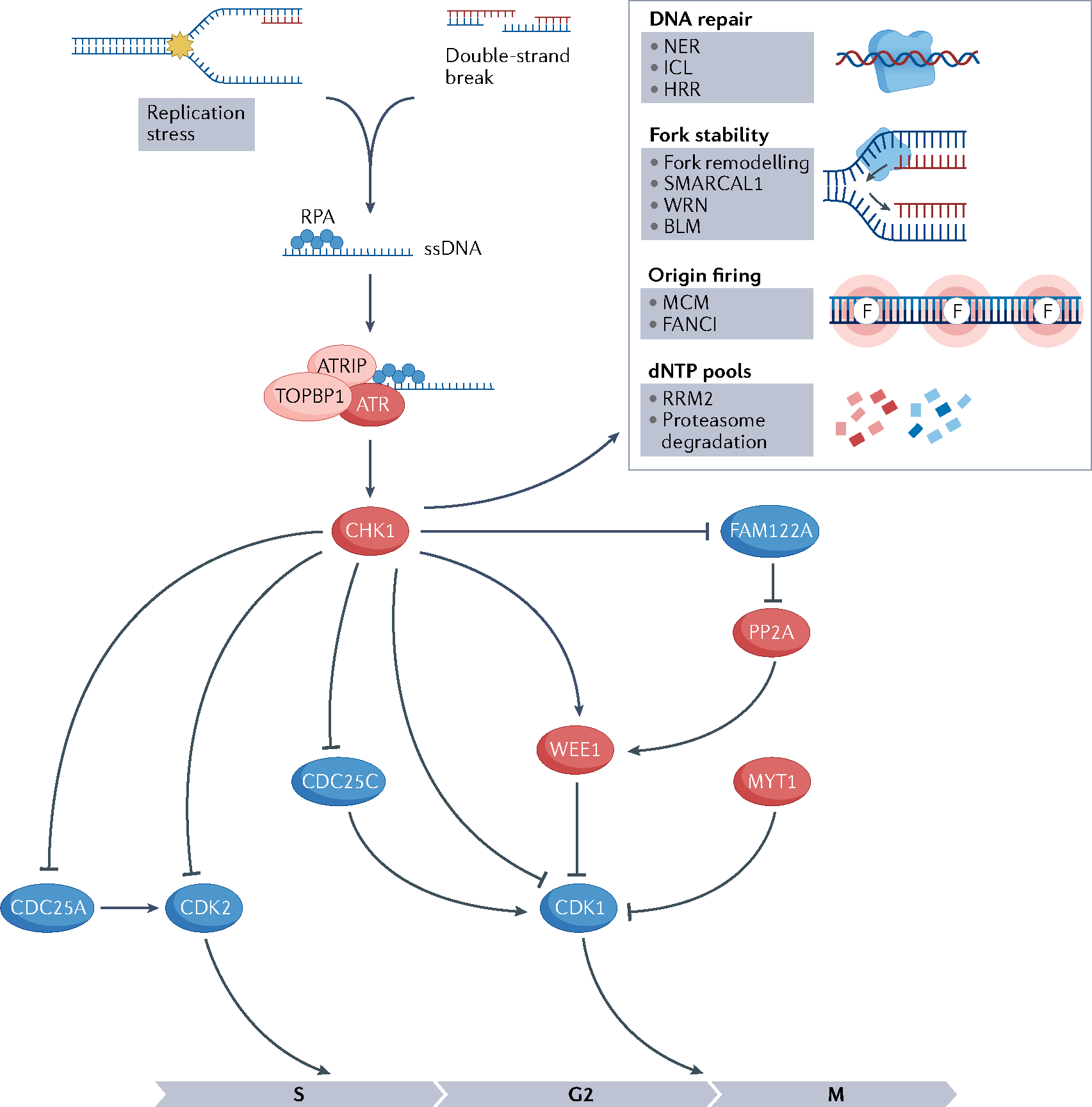
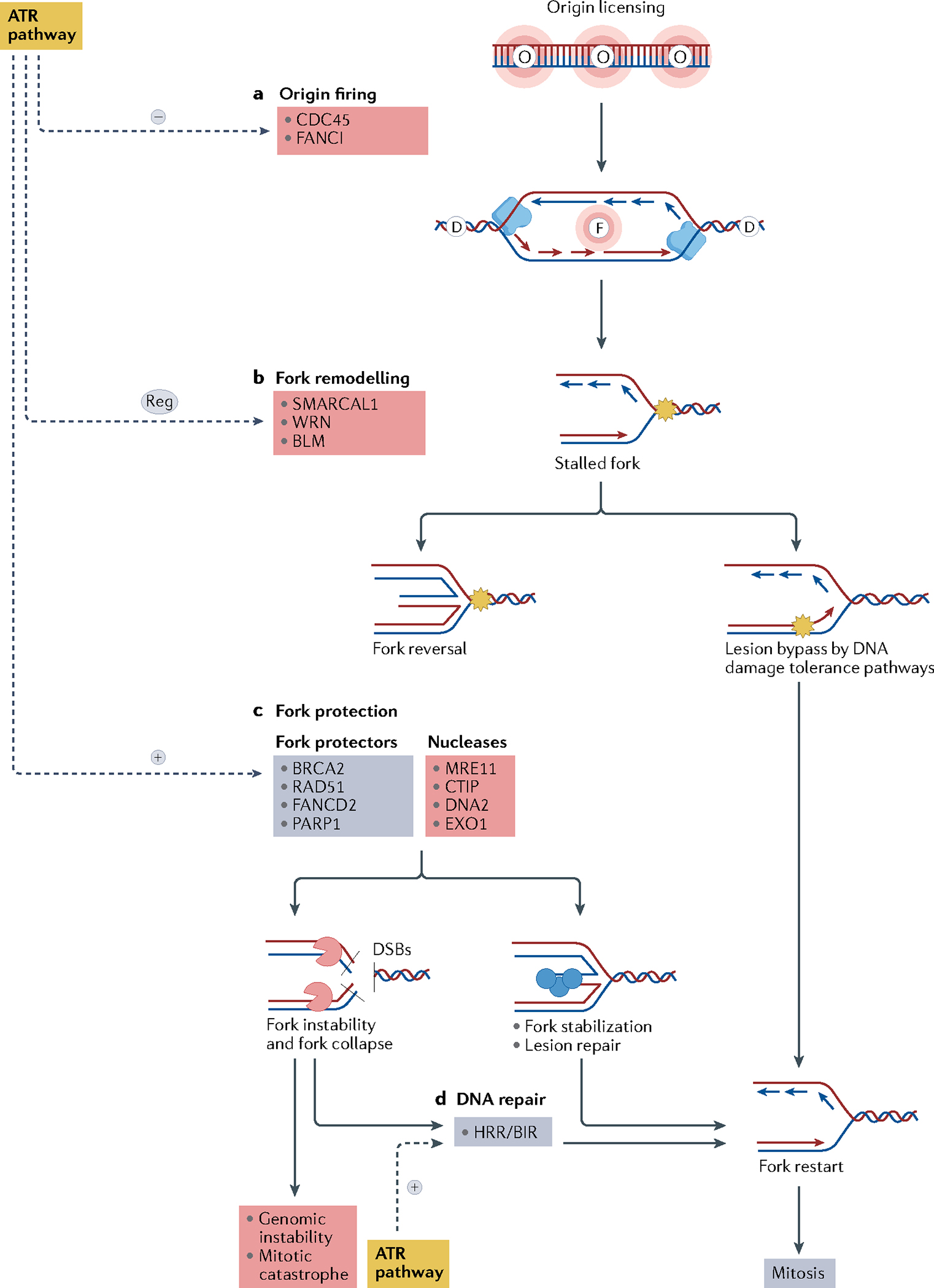
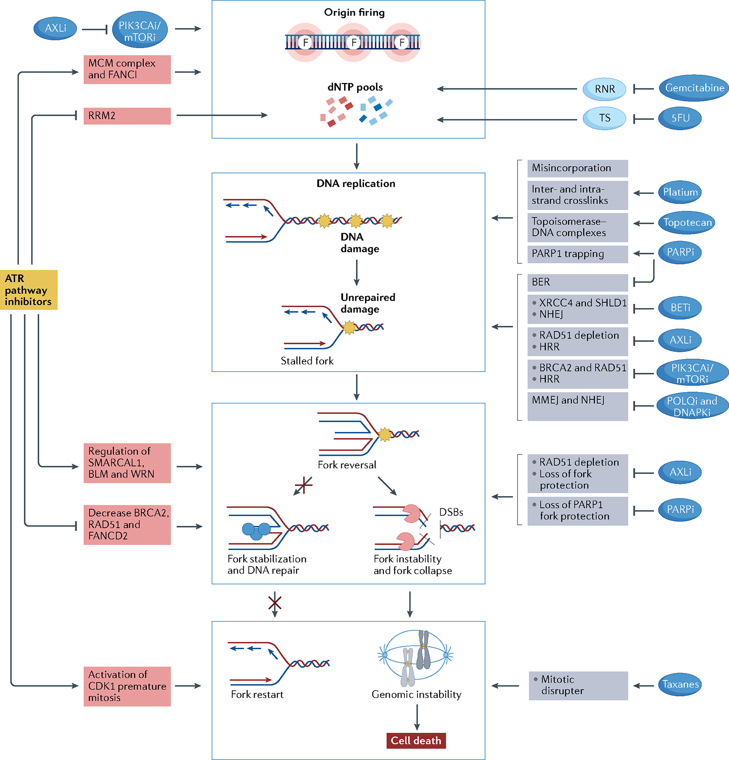
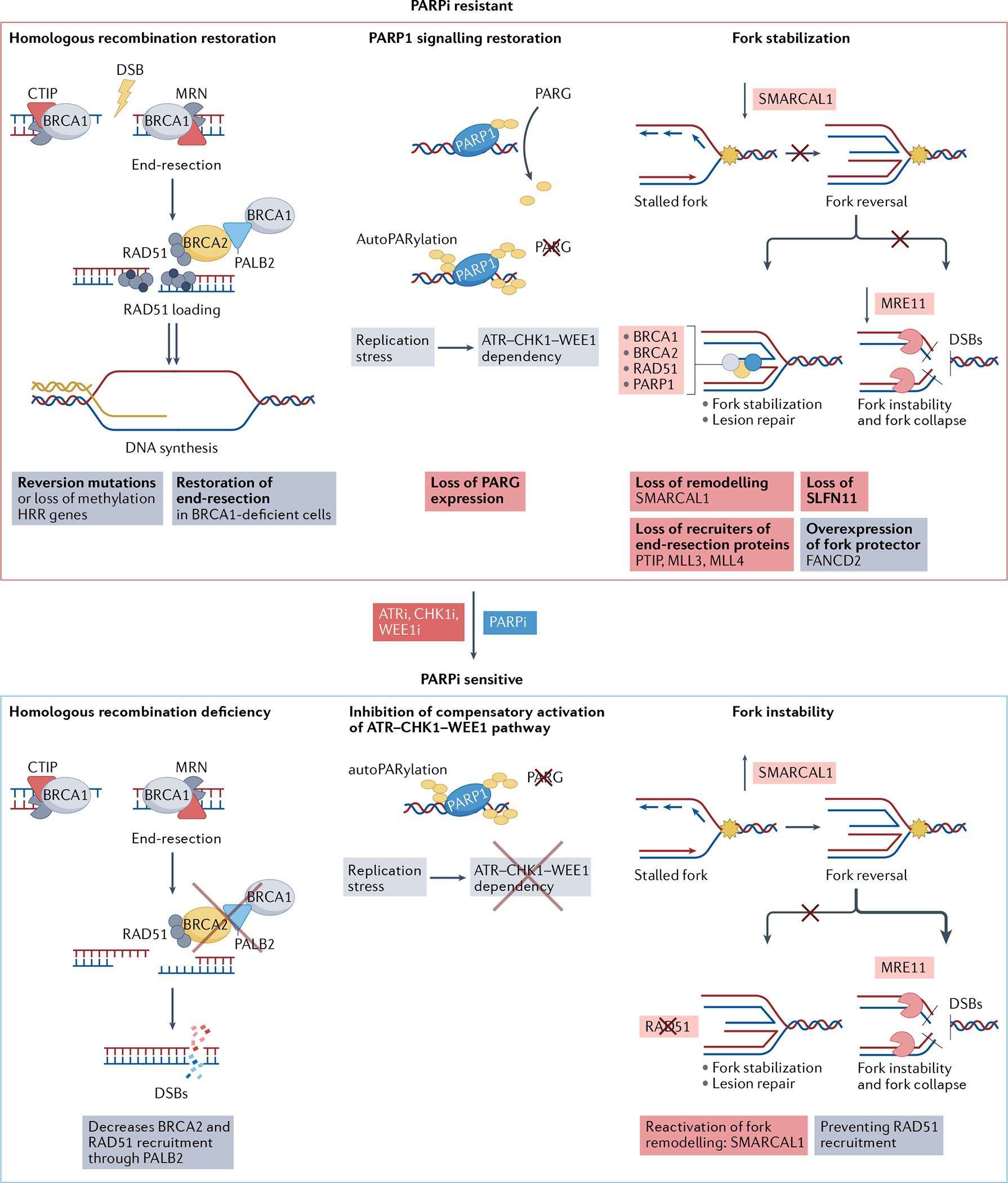
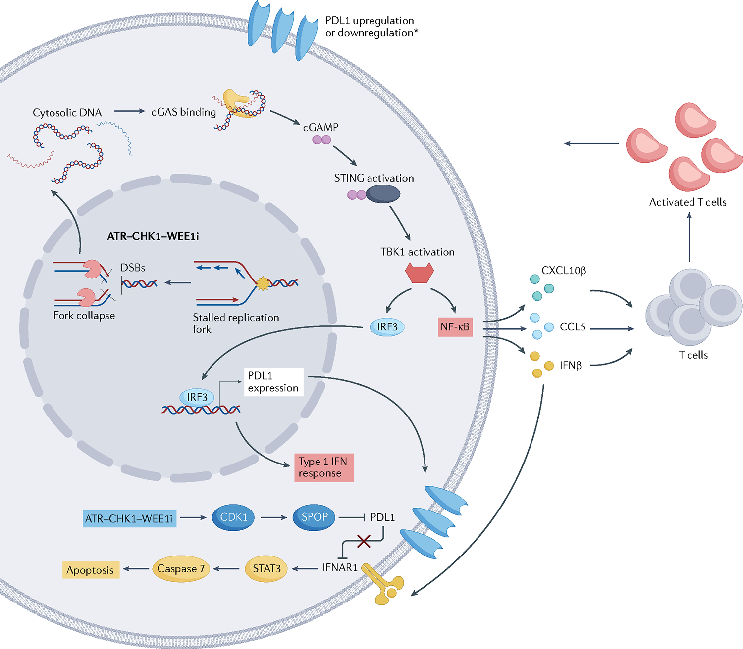
Replication stress is a major cause of genomic instability and a crucial vulnerability of cancer cells. This vulnerability can be therapeutically targeted by inhibiting kinases that coordinate the DNA damage response with cell cycle control, including ATR, CHK1, WEE1 and MYT1 checkpoint kinases. In addition, inhibiting the DNA damage response releases DNA fragments into the cytoplasm, eliciting an innate immune response. Therefore, several ATR, CHK1, WEE1 and MYT1 inhibitors are undergoing clinical evaluation as monotherapies or in combination with chemotherapy, poly[ADP-ribose]polymerase (PARP) inhibitors, or immune checkpoint inhibitors to capitalize on high replication stress, overcome therapeutic resistance and promote effective antitumour immunity. Here, we review current and emerging approaches for targeting replication stress in cancer, from preclinical and biomarker development to clinical trial evaluation.


Conflict of interest statement

Competing interests

G.I.S. is a consultant/advisory board member for Lilly, Sierra Oncology, Merck-EMD Serono, Pfizer, Astex, Almac, Roche, Bicycle Therapeutics, Fusion Pharmaceuticals, G1 Therapeutics, Bayer, Ip-sen, Cybrexa Therapeutics, Angiex, Daiichi Sankyo and Seattle Genetics, and reports receipt of commercial research grants from Lilly, Sierra Oncology, Merck-EMD Serono and Merck & Co. P.A.K. reports participation in advisory boards from GlaxoSmithKline/Tesaro, Merck, AstraZeneca and Bayer. A.D.D. is a consultant and/or advisory board member for AstraZeneca, Bayer AG, Cedilla Therapeutics, Celgene, Cyteir Therapeutics, Epizyme, GalaxoSmithKline, Ideaya, Impact Therapeutics, LAV Global Management Company Limited. D.C. and A.A.B.A.C. declare no competing interests.

Figures

Fig. 1 |
Fig. 1 |. Illustration of causes of replication stress.
A replication fork consists of an enzyme with helicase activity (shown in yellow) and an enzyme with polymerase activity (shown in blue). Replicative stress results from endogenous or exogenous obstacles to DNA replication. The red arrows indicate the direction of the continuous DNA synthesis on the leading strand and the blue arrows indicate the direction of the non-continuous DNA synthesis on the lagging strand. Oncogenic activation and some chemotherapeutic agents such as gemcitabine cause depletion of deoxynucleotides (dNTPs), which impairs the progression of ongoing DNA replication. Increased origin firing (shown by pink circles labelled with the letter F) may be caused by oncogenic activation or loss of tumour suppressor genes. Different types of DNA damage caused by endogenous and exogenous agents lead to replication stress if left unrepaired. Repetitive DNA sequence, rehybridization of the nascent RNA to DNA, forming R-loops, misincorporation of ribonucleotides and secondary DNA structures such as hairpins and quadruplexes pose a challenge to DNA replication and are endogenous causes of replication stress even in healthy cells. Finally, collisions between replication and transcription machinery result in replication stress. RNA polymerase is shown as the blue protein and the newly synthesized RNA molecule is shown in purple.
Fig. 2 |
Fig. 2 |. Schematics of the ATR pathway.
Activation of ataxia telangiectasia mutated (ATM) and Rad3-related (ATR) pathway because of replication stress or double-strand breaks results in cell cycle arrest, activation of DNA repair pathways, fork stabilization, inhibition of origin firing, decrease in deoxynucleotide (dNTP) degradation and increase in dNTP synthesis. Barriers to replication fork progression (shown by the yellow star) lead to the uncoupling of helicases and polymerases, generating single-stranded DNA (ssDNA) at the stalled fork. Replication protein A (RPA) binds to ssDNA at the stalled fork and recruits ATR through the partner ATR-interacting protein (ATRIP). ATR is then activated by DNA topoisomerase 2-binding protein 1 (TOPBP1), activates CHK1 through phosphorylation and triggers the ATR–CHK1 pathway. CHK1 mediates cell cycle arrest in the S–G2 phase through phosphorylation and reduction of cyclin-dependent kinase 2 (CDK2) activation. CDK1 is negatively regulated via phosphorylation by WEE1 and MYT1 kinases and positively regulated by CDC25 phosphatases. CHK1 phosphorylates and inactivates CDC25C, activates WEE1 and promotes the degradation of CDC25A,. FAM122A is a negative regulator of the G2/M checkpoint and forms a complex with the phosphatase PP2A. When CHK1 phosphorylates FAM122A, PP2A is disinhibited and dephosphorylates WEE1, preventing its degradation and activating the G2/M checkpoint by WEE1. Proteins whose activity leads to cell cycle arrest are shown in red and proteins whose activity leads to cell cycle progression are shown in blue. Red DNA strands indicate newly synthesized DNA. Pointed arrows indicate action on that protein (phosphorylation) and blunted arrows indicate inhibition. BLM, Bloom syndrome protein; HRR, homologous recombination repair pathway; ICL, inter-strand crosslink pathway; MCM, minichromosome maintenance; NER, nucleotide excision repair pathway; RRM2, ribonucleoside-diphosphate reductase subunit M2; WRN, Werner syndrome ATP-dependent helicase.
Fig. 3 |
Fig. 3 |. Fork dynamics regulation by the ATR pathway.
a | The ataxia telangiectasia mutated (ATM) and Rad3-related (ATR) pathway inhibits replication fork origin firing through inhibition of the minichromosome maintenance 2–7 complex (MCM2–7) and Fanconi anaemia group I protein (FANCI). Replication origin firing is indicated by the letter F, and dormant replication origins — licensed but not activated — are indicated by the letter D. Replisome (blue), is a protein complex with helicase and polymerase activities that progresses bidirectionally, starting from the origin firing and forming the replication forks. b | Stalled forks owing to an obstacle to fork progression (shown by the yellow star) may be rescued directly by DNA damage tolerance pathways or may be reversed by fork remodelling proteins. Fork reversal may result in fork protection or fork degradation depending on the balance of activity of various remodelling factors and on the DNA repair proficiency of the cell. c | The ATR pathway modulates fork reversal and activates fork protectors, promoting fork stabilization. Reversed forks are substrates for nucleases that cleave the DNA to form double-strand breaks (DSBs). Fork protectors such as BRCA2, RAD51 and poly[ADP-ribose]polymerase 1 (PARP1) prevent fork degradation by nucleases, allowing lesion repair and fork restart. d | Collapsed replication forks may be repaired by the homologous recombination repair (HRR) and the break-induced replication (BIR) pathways, allowing fork restart and normal mitosis. If DNA repair is not effective, fork collapse may lead to genomic instability and mitotic catastrophe. Mechanisms that lead to fork stabilization and restart and normal mitosis are shown in blue, mechanisms that lead to fork instability, fork collapse and genomic instability are shown in red. BLM, Bloom syndrome protein; CDC45, cell division cycle 45; WRN, Werner syndrome ATP-dependent helicase.
Fig. 4 |
Fig. 4 |. Drug combinations with ATR–CHK1–WEE1 inhibitors.
Rationale for drug combinations that promote replication stress, potentiating the effect of ATR–CHK1–WEE1 inhibitors. Red boxes indicate mechanisms of action related to ataxia telangiectasia mutated (ATM) and Rad3-related (ATR) pathway inhibitors; grey boxes indicate mechanisms of action of the other drugs. Central blue squares group the main mechanisms through which drugs may activate replication stress. Drugs may activate replication stress by decreasing deoxynucleotide (dNTP) pools directly or through increasing origin firing (indicated by the letter F, top blue box), increasing DNA damage (second blue box), promoting fork instability (third blue box) and disrupting mitosis (bottom blue box). ATR inhibitors (ATRi), CHK1 inhibitors (CHK1i) and WEE1 inhibitors (WEE1i) decrease dNTP pools by activating origin firing through loss of Fanconi anaemia group I protein (FANCI) phosphorylation and minichromosome maintenance 2–7 complex (MCM2–7) inhibition and by decreasing ribonucleoside-diphosphate reductase subunit M2 (RRM2) expression. PIK3CA/mTOR inhibitors synergize with ATRi, CHK1i or WEE1i by promoting origin firing through increasing cell division cycle 45 (CDC45) expression, which leads to activation of the MCM2–7 complex. Moreover, AXL inhibitors (AXLi) decrease dNTP pools by inhibiting the PI3K–mTOR pathway. Nucleoside analogues reduce intracellular dNTP pools by inhibiting the enzymes responsible for dNTP synthesis. Gemcitabine inhibits ribonucleotide reductase (RNR), and 5-fluorouracil (5FU) inhibits thymidylate synthetase (TS). Drugs may increase DNA damage by directly damaging the DNA or by inhibiting DNA repair mechanisms. Gemcitabine and 5FU lead to DNA damage by their misincorporation into DNA. Platinum salts cause inter- and intra-strand crosslinks, and topotecan generates topoisomerase–DNA complexes, which become obstacles to fork progression. Poly[ADP-ribose]polymerase (PARP) inhibitors (PARPi) create obstacles to fork progression through PARP1 trapping and inhibition of the base excision repair (BER) pathway. Bromodomain and extra-terminal domain (BET) inhibitors (BETi) lead to non-homologous end-joining (NHEJ) inhibition through downregulation of XRCC4 and SHLD1. AXL inhibition results in inhibition of homologous recombination repair (HRR) through RAD51 depletion. PIK3CA/mTOR inhibition leads to HRR inhibition through downregulation of BRCA2 and RAD51. POLQ inhibitors and DNAPK inhibitors directly inhibit microhomology-mediated end-joining (MMEJ) and NHEJ pathways, respectively. ATRi, CHK1i and WEE1i cause fork instability by regulating fork remodellers such as SMARCAL1, Bloom syndrome protein (BLM) and Werner syndrome ATP-dependent helicase (WRN) and by decreasing the activity of fork protectors such as BRCA2, RAD51 and FANCD2. AXLi and PARPi also lead to fork instability by depletion of RAD51 and inhibition of PARP1 fork protection, respectively. Finally, ATRi, CHK1i and WEE1i lead cells to premature mitosis and may synergize with mitotic disrupter agents such as taxanes.
Fig. 5 |
Fig. 5 |. Mechanisms of ATR–CHK1–WEE1 inhibitors to overcome PARP inhibitor resistance.
The top panel shows mechanisms of poly[ADP-ribose]polymerase (PARP) inhibitor (PARPi) resistance. The main steps of the homologous recombination repair (HRR) pathway are shown at the left: DNA end resection on double-strand breaks (DSBs), RAD51 loading, homologous strand invasion and DNA synthesis. HRR deficiency makes cells sensitive to PARPi. Reversion mutations in BRCA1 and BRCA2, and RAD51 paralogues can restore HRR. CtIP and MRN are nucleases responsible for initial DNA end resection. MRN is a nuclease complex formed by MRE11-RAD50-NBS1. Loss of proteins that inhibit DNA end resection, such as 53BP1 and shieldin complex proteins, or upregulation of proteins that promote end resection, such as TRIP13 and USP15, can restore end resection in BRCA1-deficient cells, promoting PARPi resistance. The central panel shows how poly(ADP-ribose) glycohydrolase (PARG) counteracts PARP1-mediated PARylation (shown in yellow). Accordingly, loss of expression of PARG can lead to upregulation of PAR despite PARP1 inhibition, increasing replication stress and activating the ATR–CHK1–WEE1 pathway. The top right panel shows a stalled fork undergoing fork reversal. Fork protectors (shown by the grey, yellow and blue circles) such as BRCA1, BRCA2, RAD51 and PARP1 stabilize the fork, allowing DNA repair and fork restart. In the absence of fork protectors, nucleases (shown in red) such as MRE11 can degrade the reversed forks. Accordingly, loss of fork remodelling factors such as SMARCAL1, loss of recruiters of end resection such as PTIP, MLL3 and MLL4, and overexpression of fork protectors such as Fanconi anaemia group D2 protein (FANCD2), can lead to fork stabilization and PARPi resistance. The bottom panels show how ataxia telangiectasia mutated (ATM) and Rad3-related inhibitors (ATRi), CHK1 inhibitors (CHK1i) and WEE1 inhibitors (WEE1i) can overcome PARPi resistance. ATRi, CHK1i and WEE1i can cause HRR deficiency by decreasing BRCA2 and RAD51 recruitment (bottom left panel). They can suppress activation of the ATR pathway, which may be overactivated by PARG loss (bottom centre panel), and promote fork instability by reactivating SMARCAL1 and preventing RAD51 recruitment to the reversed fork (bottom right panel). SLFN11, Schlafen family member 11.
Fig. 6 |
Fig. 6 |. Mechanism of immune response sensitization.
Replication stress can elicit an innate immune response by releasing DNA particles into the cytoplasm, activating the cGAS–stimulator of interferon genes (STING) pathway, and upregulating the type I interferon-γ (IFNγ) response and, subsequently, the nuclear factor-κB (NF-κB) pathway. Besides this pro-inflammatory signal, cGAS–STING can also upregulate PDL1 expression. In addition, the ATR–CHK1 pathway can also lead to PDL1 protein degradation by activating the CDK1–speckle-type POZ protein (SPOP) pathway. Downregulation of PDL1 can inhibit an IFNβ-mediated pro-apoptotic pathway, resulting in autocrine cytotoxic signalling. IFNAR1, interferon-α/β receptor 1. *The resulting effect of ATR-CHK1-WEE1 inhibition on PD-L1 is disputable with studies showing both upregulation or downregulation upon blockage of the ATR-CHK1-WEE1 pathway.

References

    1. Hanahan D & Weinberg RA Hallmarks of cancer: the next generation. Cell 144, 646–674 (2011). - PubMed
    1. Zeman MK & Cimprich KA Causes and consequences of replication stress. Nat. Cell Biol. 16, 2–9 (2014). - PMC - PubMed
    1. Gaillard H, García-Muse T & Aguilera A Replication stress and cancer. Nat. Rev. Cancer 15, 276–280 (2015). - PubMed
    1. Halazonetis TD, Gorgoulis VG & Bartek J An oncogene-induced DNA damage model for cancer development. Science 319, 1352–1355 (2008). - PubMed
    1. Wilhelm T, Said M & Naim V DNA replication stress and chromosomal instability: dangerous liaisons. Genes 11, 1–35 (2020). - PMC - PubMed

Publication types