Review
. 2023 Feb;396(2):173-190.
doi: 10.1007/s00210-022-02300-9. Epub 2022 Oct 7.

From signal transduction to protein toxins-a narrative review about milestones on the research route of C. difficile toxins

Affiliations
Review

From signal transduction to protein toxins-a narrative review about milestones on the research route of C. difficile toxins

Klaus Aktories. Naunyn Schmiedebergs Arch Pharmacol. 2023 Feb.

Abstract

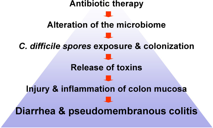
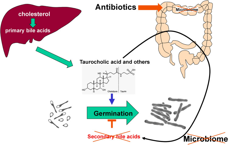
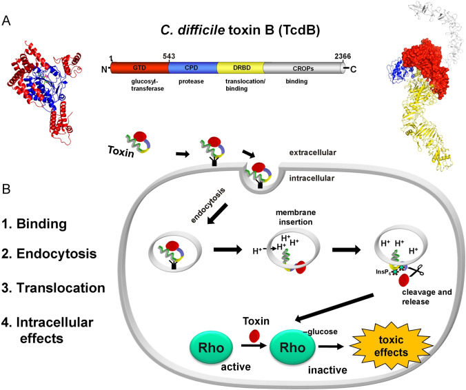
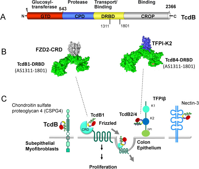
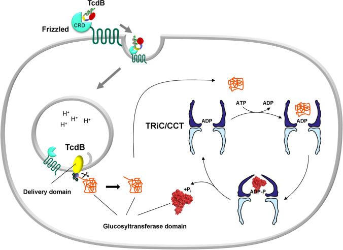
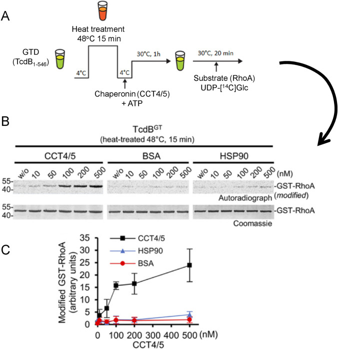
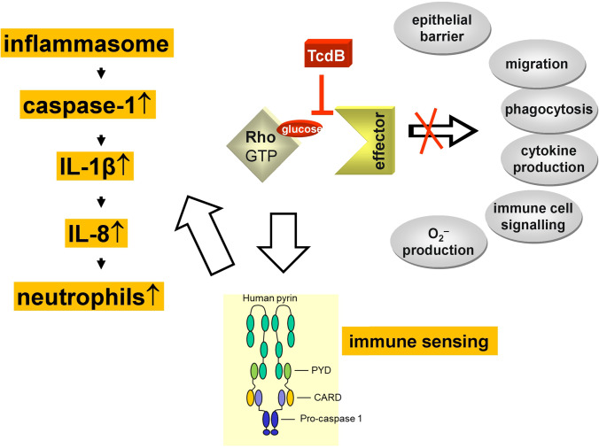
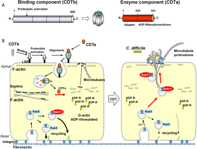
Selected findings about Clostridioides difficile (formerly Clostridium difficile) toxins are presented in a narrative review. Starting with a personal view on research about G proteins, adenylyl cyclase, and ADP-ribosylating toxins in the laboratory of Günter Schultz in Heidelberg, milestones of C. difficile toxin research are presented with the focus on toxin B (TcdB), covering toxin structure, receptor binding, toxin up-take and refolding, the intracellular actions of TcdB, and the treatment of C. difficile infection.

Keywords: Bacterial protein toxins; C. difficile ADP-ribosyltransferase CDT; C. difficile toxin TcdB; G proteins; Pseudomembranous colitis; Toxin receptors; Toxin up-take.


Conflict of interest statement

The author declares no competing interests.

The author declares no competing interests.

Figures

Fig. 1
Fig. 1
Antibiotics-induced C. difficile infection (CDI). The various steps involved in CDI are exhibited
Fig. 2
Fig. 2
Effects of bile acids on germination of C. difficile spores. Germination of C. difficile spores is enhanced by primary bile acids and bile acid conjugates (like taurocholic acid). Secondary bile acids, which are produced from primary bile acids by the gut microbiome, inhibit germination. Therefore, antibiotics, which damage the gut microbiome, enhance germination of spores
Fig. 3
Fig. 3
C. difficile toxin B structure and action. A. Left part: the structure of the glucosyltransferase domain of C. difficile toxin B (TcdB) is shown (In dark blue, is the catalytic core of the glucosyltransferase depicted and in red peripheral helices are given. Middle part: Scheme of the 4 functional domains of TcdB. At the N-terminus is the glucosyltransferase domain (GTD, red), it follows the inherent cysteine protease (blue), the delivery and binding domain (DRBD, yellow) and at the C-terminus the CROPs domain (gray), which is also involved in binding. Right part: Crystal structure of the complete TcdB. Domains are colored as in the linear scheme depicted in the middle part. B. left part: The typical steps of the actions of an intracellularly acting toxin are listed. Right part: The toxin (e.g., TcdB) binds to its receptor and is endocytosed, at low pH of endosomes the toxin is able to insert into the endosomal membrane. In the cytosol, the cysteine protease domain is activated by InsP6 (inositol hexakisphosphate), thereby the glucosyltransferase domain (GTD) is released. GTD modifies Rho proteins by mono-O-glucosylation and blocks the regulatory actions of these switch proteins. Pictures were designed using (10.2210/pdb7v1n/pdb) and (10.2210/pdb2BVL/pdb) by PyMol
Fig. 4
Fig. 4
Receptors and binding of TcdB. A. Schema of the 4 domain structure of TcdB. the binding region for receptor interaction with the receptors frizzled (FZD) and tissue factor pathway inhibitor (TFPI) is indicated (residues 1311–1801). B. Surface view of the crystal structure of part of the delivery/receptor-binding domain (DRBD) of TcdB in complex with the receptors FZD and TFPI. The toxin subtype TcdB1 binds to the cysteine rich domain of frizzled (FZD1-CRD), while the toxin subtype TcdB4 binds to the Kunitz domain 2 of TFPI (TFPI-K2). The binding areal of both toxin subtypes is almost identical. C. Various receptors of TcdB are shown. Chrondroitin sulfate glycoprotein 4 (CSPG4) binds both toxin subtypes but is not located on the intestinal epithelium. The heptahelical frizzled receptor binds TcdB1 with its cysteine rich domain (CRD). Frizzled is a receptor for Wnt ligand and involved in proliferation. TFPI binds with its Kunitz 2 domain TcdB4. Nectin-3 has been also identified as TcdB receptor. Its pathophysiological role is not clear. Picture was designed on the bases of (10.2210/pdb6C0B/pdb) and (10.2210/pdb7V1N/pdb) by PyMol
Fig. 5
Fig. 5
Model of the action of the chaperonin TriC/CCT in TcdB up-take and action. TcdB binds to its receptor Frizzled and is endocytosed. At low pH of endosomes at least GTD and CPD od TcdB are translocated into the cytosol. Most likely, translocation occurs as a single chain. In the cytosol, GTD is refolded with the help of the chaperonin TriC/CCT. The chaperonin has a double cage-like structure and itsaction depends on hydrolysis of ATP. Schema modified from (Russmann et al. 2012)
Fig. 6
Fig. 6
Involvement of chaperonin TriC/CCT in stabilization and refolding of the glucosyltransferase domain of TcdB. A. The experimental procedure to show the role of chaperonin is given. The glucosyltransferase domain GTD (residues 1–546) was heated for 15 min at 48 °C, then the chaperonin (CCT4/5) was added together with ATP and the mixture remained for 1 h at 30 °C. Then, the glucosyltransferase activity was studied by addition of the substrate RhoA and radioactively labeled UDP-glucose as a sugar donor. B. The autoradiograph shows that CCT4/5 stabilized (refolded) GTD in a concentration-dependent manner. BSA or the HSP90 chaperone were without effects. C. Quantification and statistics of the experiment given under B. (Data from Steinemann et al. 2018)
Fig. 7
Fig. 7
Functional consequences of Rho inactivation by TcdB-induced glucosylation. Glucosylation of Rho inhibits the interaction of this switch protein with numerous effectors, thereby epithelial barrier functions, cell migration, phagocytosis, cytokine production, immune cell signaling and O2− production is blocked. On the other hand, Rho inhibition causes pyrine inflammasome activation, eventually resulting in release of IL-1β with subsequent IL-8 release and attraction of neutrophil leukocytes
Fig. 8
Fig. 8
Structure and actions of C. difficile ADP-ribosyltransferase CDT. A. CDT is a binary toxin and consists of two separated toxin components, the binding component CDTb and the enzyme component CDTa. CDTb is proteolytically activated and forms heptamers. CDTa has an adaptor domain at the N-terminus and an enzyme domain (ADP-ribosyltransferase) at the C-terminus. B. Model of the actions of CDT. Left cell: CDTb binds to LSR, is proteolytically activated and forms hepatmers. So far it is unclear, whether the activation step is before or after receptor binding. The heptamers bind the enzyme component CDTa. The receptor-toxin complex is endocytosed. At low pH of endosomes CDTb forms pores and translocates CDTa into the cytosol. Here, CDTa ADP-ribosylated G-actin and inhibits the polymerisation of actin. Depolymerisation of submembranous F-actin allows formation of microtubule-based protrusions and releases septins from F-actin, which guide the microtubules into the protrusions. The lower part shows the recycling of vesicles with integrin and bound fibronectin. Right cell: CDTa-induced ADP-ribosylation of actin results in misguiding of vesicles (Rab11-associated vesicles) to the apical membrane, where fibronectin is released. Microtubule-based protrusions and fibronectin enhance binding of C. difficile bacteria
Fig. 9
Fig. 9
Effects of C. difficile ADP-ribosyltransferase CDT on Caco-2 cells. A. Addition of CDT to Caco-2 cell culture results in formation of long microtubule-based cell protrusion (left, control; right CDT). B. The net, formed by microtubule-based cell protrusions, increases the adherence of C. difficile bacteria C. Septins (yellow and arrow head) are involved in guiding of microtubles at the membrane. Septins form a funnel-like structure for microtubles. Data from Schwan et al. and from Nölke et al., 2016
Fig. 10
Fig. 10
Treatment options for diseases caused by C. difficile infection. Colored ovates represent approved treatments including the antibiotics fidaxomicin, vancomycin, metronidazole, and tigecycline, the anti-TcdB-antibody bezlotoxumab and fecal microbiota transplantation (FMT). The dotted ovates show proposed future treatment options (for details see text). The structure of TcdB shows the target sites of Bezlotoxumab, the binding sites of three neutralizing monovalent antibody E3, 7F, 5D and the binding region of FZD and TFPI

References

    1. Abt MC, McKenney PT, Pamer EG. Clostridium difficile colitis: pathogenesis and host defence. Nat Rev Microbiol. 2016;14:609–620. doi: 10.1038/nrmicro.2016.108. - DOI - PMC - PubMed
    1. Aktories K. Bacterial protein toxins that modify host regulatory GTPases. Nat Rev Microbiol. 2011;9:487–498. doi: 10.1038/nrmicro2592. - DOI - PubMed
    1. Aktories K, Hall A. Botulinum ADP-ribosyltransferase C3: a new tool to study low molecular weight GTP-binding proteins. Tips. 1989;10:415–418. - PubMed
    1. Aktories K, Bärmann M, Ohishi I, Tsuyama S, Jakobs KH, Habermann E. Botulinum C2 toxin ADP-ribosylates actin. Nature. 1986;322:390–392. doi: 10.1038/322390a0. - DOI - PubMed
    1. Aktories K, Weller U, Chhatwal GS. Clostridium botulinum type C produces a novel ADP- ribosyltransferase distinct from botulinum C2 toxin. FEBS Lett. 1987;212:109–113. doi: 10.1016/0014-5793(87)81566-1. - DOI - PubMed

Publication types

MeSH terms

Substances

LinkOut - more resources