Review
. 2023 May;38(5):1411-1425.
doi: 10.1007/s00467-022-05755-3. Epub 2022 Oct 6.

The complement system in pediatric acute kidney injury

Affiliations
Review

The complement system in pediatric acute kidney injury

Erin K Stenson et al. Pediatr Nephrol. 2023 May.

Abstract

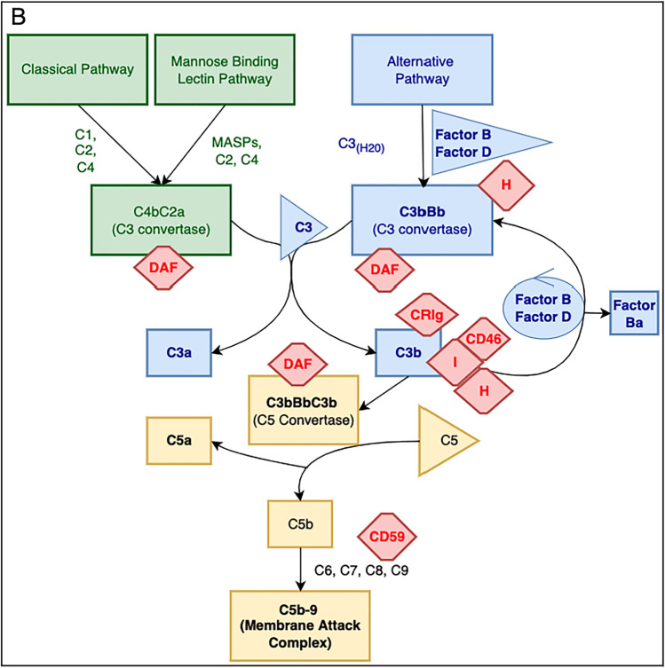
The complement cascade is an important part of the innate immune system. In addition to helping the body to eliminate pathogens, however, complement activation also contributes to the pathogenesis of a wide range of kidney diseases. Recent work has revealed that uncontrolled complement activation is the key driver of several rare kidney diseases in children, including atypical hemolytic uremic syndrome and C3 glomerulopathy. In addition, a growing body of literature has implicated complement in the pathogenesis of more common kidney diseases, including acute kidney injury (AKI). Complement-targeted therapeutics are in use for a variety of diseases, and an increasing number of therapeutic agents are under development. With the implication of complement in the pathogenesis of AKI, complement-targeted therapeutics could be trialed to prevent or treat this condition. In this review, we discuss the evidence that the complement system is activated in pediatric patients with AKI, and we review the role of complement proteins as biomarkers and therapeutic targets in patients with AKI.

Keywords: Acute kidney injury; Complement; Complement inhibitors.


Conflict of interest statement

Dr. Stenson and Dr. Kendrick declare they have no financial interests. Dr. Dixon is a consultant for Apellis and Alexion Pharmaceuticals, Inc. Dr. Thurman received royalties from Alexion Pharmaceuticals, Inc. and is a consultant for Q32 Bio, Inc., a company developing complement inhibitors. He also holds stock and will receive royalty income from Q32 Bio, Inc.

Figures

Fig. 1
Fig. 1
Overview of the complement cascade. A Activation pathways. The classical pathway, mannose-binding lectin pathway, and alternative pathway converge on C3, cleaving C3 into activation fragments C3a and C3b. C3b joins with factor B, which is then cleaved by the rate-limiting enzyme factor D. This generates the Ba fragment, which can be measured as a marker of this process. It also creates the C3 convertase (C3bBb). C3bBb is involved in the amplification loop of the alternative pathway, increasing the generation of downstream activation fragments. C3b also joins with C3bBb to create the C5 convertase (C3bBbC3b), which converts C5 into C5a and C5b. C3a and C5a function as anaphylatoxins which cause chemoattraction of myeloid cells, leukocyte activation leading to release of proinflammatory mediations, and increased vascular permeability causing vascular leak. C5b joins with C6, C7, C8, and C9 to form C5b-9, also termed the membrane attack complex (MAC) which lyses target cells. B Complement regulatory proteins. Regulatory proteins are integral in controlling the complement cascade and preventing pathologic activation within tissues. Factor H is a regulator of the alternative pathway that inactivates C3b, competes with factor B for C3b binding (and prevents formation of C3 convertase), and accelerates C3 convertase decay. Factor H is a soluble protein that controls alternative pathway activation in the fluid phase, but it can also bind to cells and extracellular matrix to control activation at those locations. CD46 is another cofactor (for factor I) that mediates inactivation of C3b. CRIg (complement receptor of immunoglobulin family) acts on C3b and inhibits alternative pathway activation. CD59 binds C8 and C9, thereby preventing the formation of the membrane attack complex C5b-9. Decay accelerating factor (DAF or CD55) increases the breakdown of the C3 and C5 convertases within the pathway
Fig. 1
Fig. 1
Overview of the complement cascade. A Activation pathways. The classical pathway, mannose-binding lectin pathway, and alternative pathway converge on C3, cleaving C3 into activation fragments C3a and C3b. C3b joins with factor B, which is then cleaved by the rate-limiting enzyme factor D. This generates the Ba fragment, which can be measured as a marker of this process. It also creates the C3 convertase (C3bBb). C3bBb is involved in the amplification loop of the alternative pathway, increasing the generation of downstream activation fragments. C3b also joins with C3bBb to create the C5 convertase (C3bBbC3b), which converts C5 into C5a and C5b. C3a and C5a function as anaphylatoxins which cause chemoattraction of myeloid cells, leukocyte activation leading to release of proinflammatory mediations, and increased vascular permeability causing vascular leak. C5b joins with C6, C7, C8, and C9 to form C5b-9, also termed the membrane attack complex (MAC) which lyses target cells. B Complement regulatory proteins. Regulatory proteins are integral in controlling the complement cascade and preventing pathologic activation within tissues. Factor H is a regulator of the alternative pathway that inactivates C3b, competes with factor B for C3b binding (and prevents formation of C3 convertase), and accelerates C3 convertase decay. Factor H is a soluble protein that controls alternative pathway activation in the fluid phase, but it can also bind to cells and extracellular matrix to control activation at those locations. CD46 is another cofactor (for factor I) that mediates inactivation of C3b. CRIg (complement receptor of immunoglobulin family) acts on C3b and inhibits alternative pathway activation. CD59 binds C8 and C9, thereby preventing the formation of the membrane attack complex C5b-9. Decay accelerating factor (DAF or CD55) increases the breakdown of the C3 and C5 convertases within the pathway
Fig. 2
Fig. 2
Site of complement activation within the renal tubulointerstitium. A Normal complement activation and regulatory control. Complement activation through any inciting pathway activates the conversion of the fluid-phase C3 into C3a and C3b. C3b deposits on the renal tubular epithelial cell in a process normally controlled by factor H and cell surface complement regulatory proteins (CRPs). B Complement activation in the setting of tubular epithelial cell injury. Stressed or injured tubular epithelial cells increase expression of L-fucose on the cell surface. Collectin-11 (CL-11) functions as a pattern recognition molecule within the mannose-binding lectin pathway. CL-11 binds to L-fucose. CL-11/L-fucose/MASP complexes then promote complement activation via cleavage of C3. C Expression of cell-surface regulatory proteins (CRPs) is disrupted after tubular epithelial cell injury. In this situation, C3b and the C3 convertase are no longer efficiently inactivated, and activation proceeds

References

    1. Hui-Stickle S, Brewer ED, Goldstein SL. Pediatric ARF epidemiology at a tertiary care center from 1999 to 2001. Am J Kidney Dis. 2005;45(1):96–101. doi: 10.1053/j.ajkd.2004.09.028. - DOI - PubMed
    1. Fitzgerald JC, Basu RK, Akcan-Arikan A, Izquierdo LM, Piñeres Olave BE, Hassinger AB, Szczepanska M, Deep A, Williams D, Sapru A, Roy JA, Nadkarni VM, Thomas N, SL JW, Furth S. Acute kidney injury in pediatric severe sepsis: an independent risk factor for death and new disability. Crit Care Med. 2016;44(12):2241–2250. doi: 10.1097/CCM.0000000000002007. - DOI - PMC - PubMed
    1. Kaddourah A, Basu RK, Bagshaw SM, Goldstein SL. Epidemiology of acute kidney injury in critically ill children and young adults. N Engl J Med. 2017;376(1):11–20. doi: 10.1056/NEJMoa1611391. - DOI - PMC - PubMed
    1. Alkandari O, Eddington KA, Hyder A, Gauvin F, Ducruet T, Gottesman R, Phan V, Zappitelli M. Acute kidney injury is an independent risk factor for pediatric intensive care unit mortality, longer length of stay and prolonged mechanical ventilation in critically ill children: a two-center retrospective cohort study. Critical Care. 2011;15(3):R146. doi: 10.1186/cc10269. - DOI - PMC - PubMed
    1. Mammen C, Al Abbas A, Skippen P, Nadel H, Levine D, Collet JP, Matsell DG. Long-term risk of CKD in children surviving episodes of acute kidney injury in the intensive care unit: a prospective cohort study. Am J Kidney Dis. 2012;59(4):523–530. doi: 10.1053/j.ajkd.2011.10.048. - DOI - PubMed

Publication types

Substances