. 2022;4(3):224-240.
doi: 10.26502/josm.511500060. Epub 2022 Sep 5.

A Pre-clinical Standard Operating Procedure for Evaluating Orthobiologics in an In Vivo Rat Spinal Fusion Model

Affiliations

A Pre-clinical Standard Operating Procedure for Evaluating Orthobiologics in an In Vivo Rat Spinal Fusion Model

Andrew L Alejo et al. J Orthop Sports Med. 2022.

Abstract

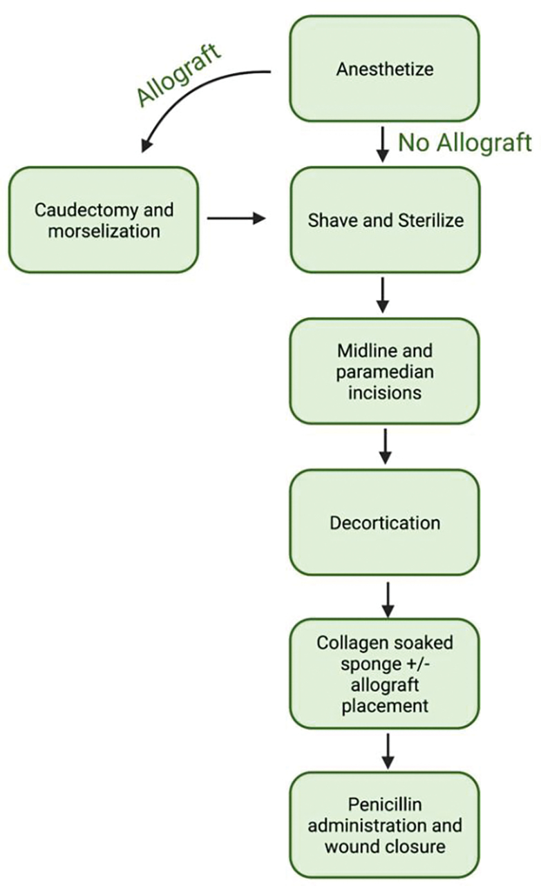
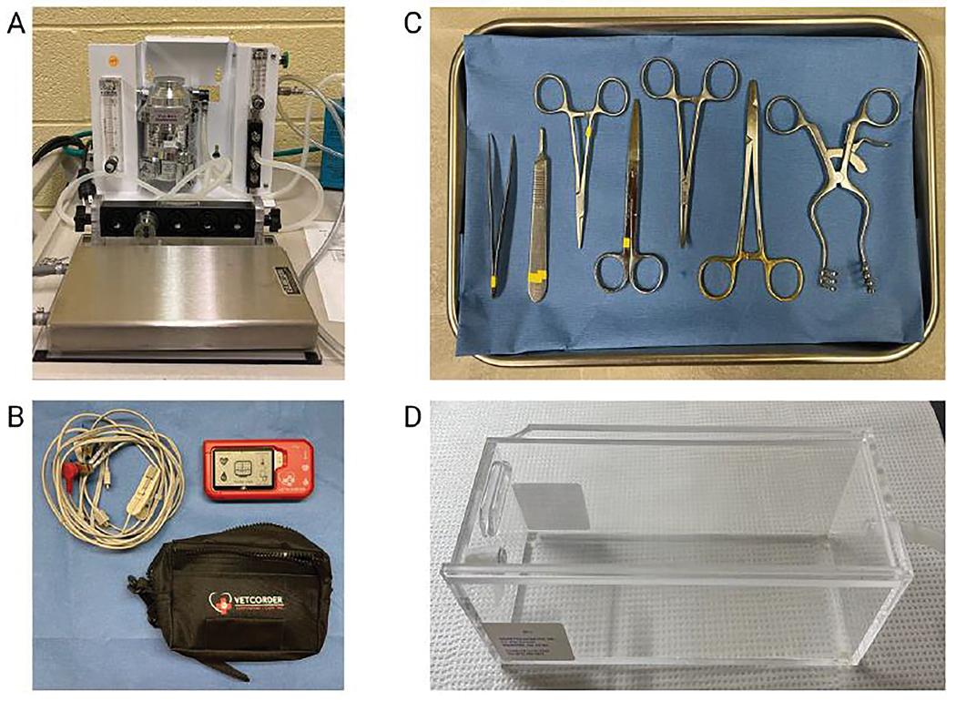
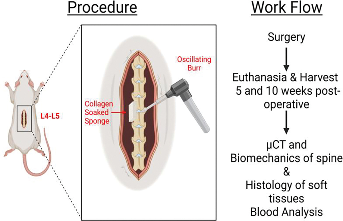
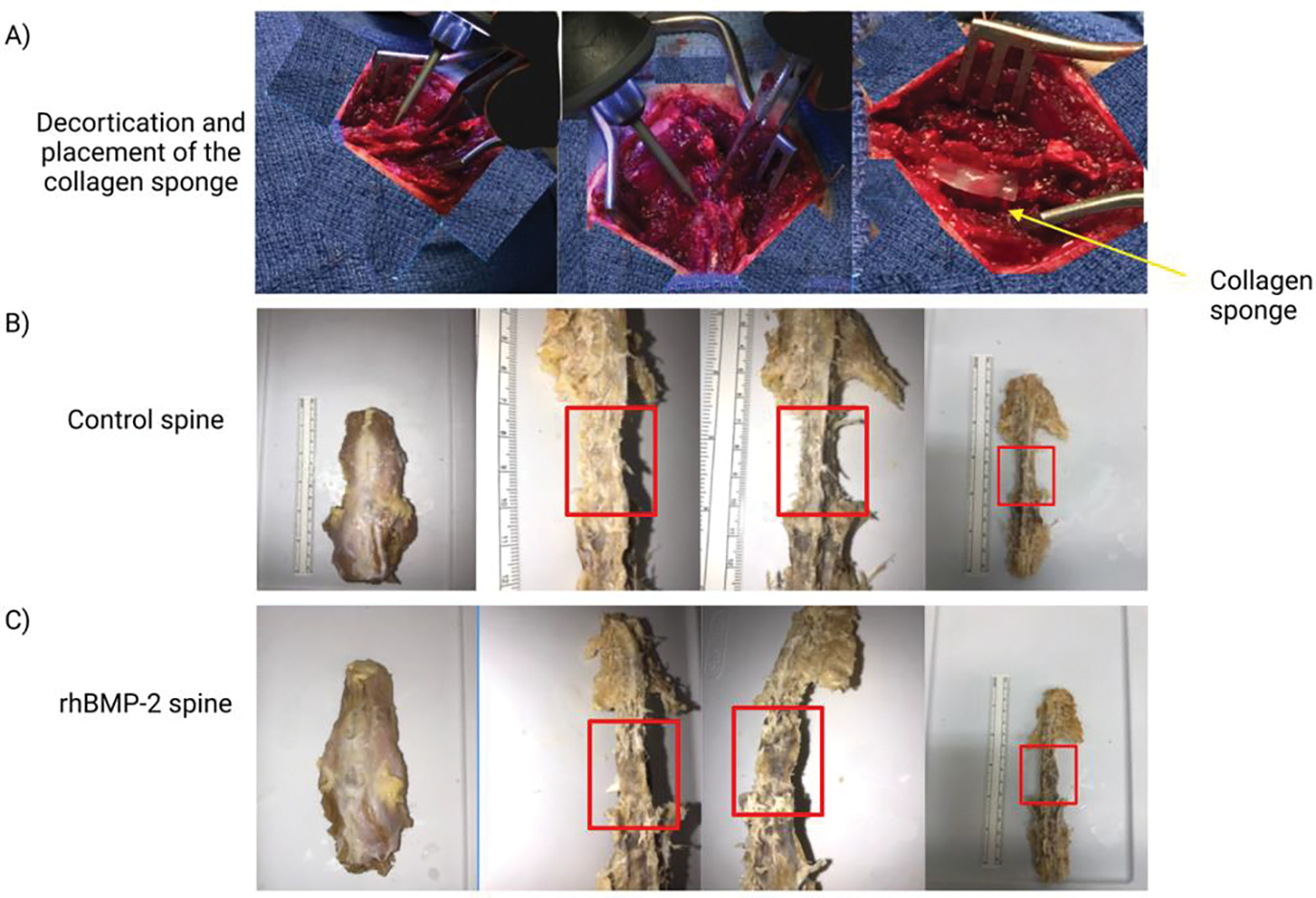
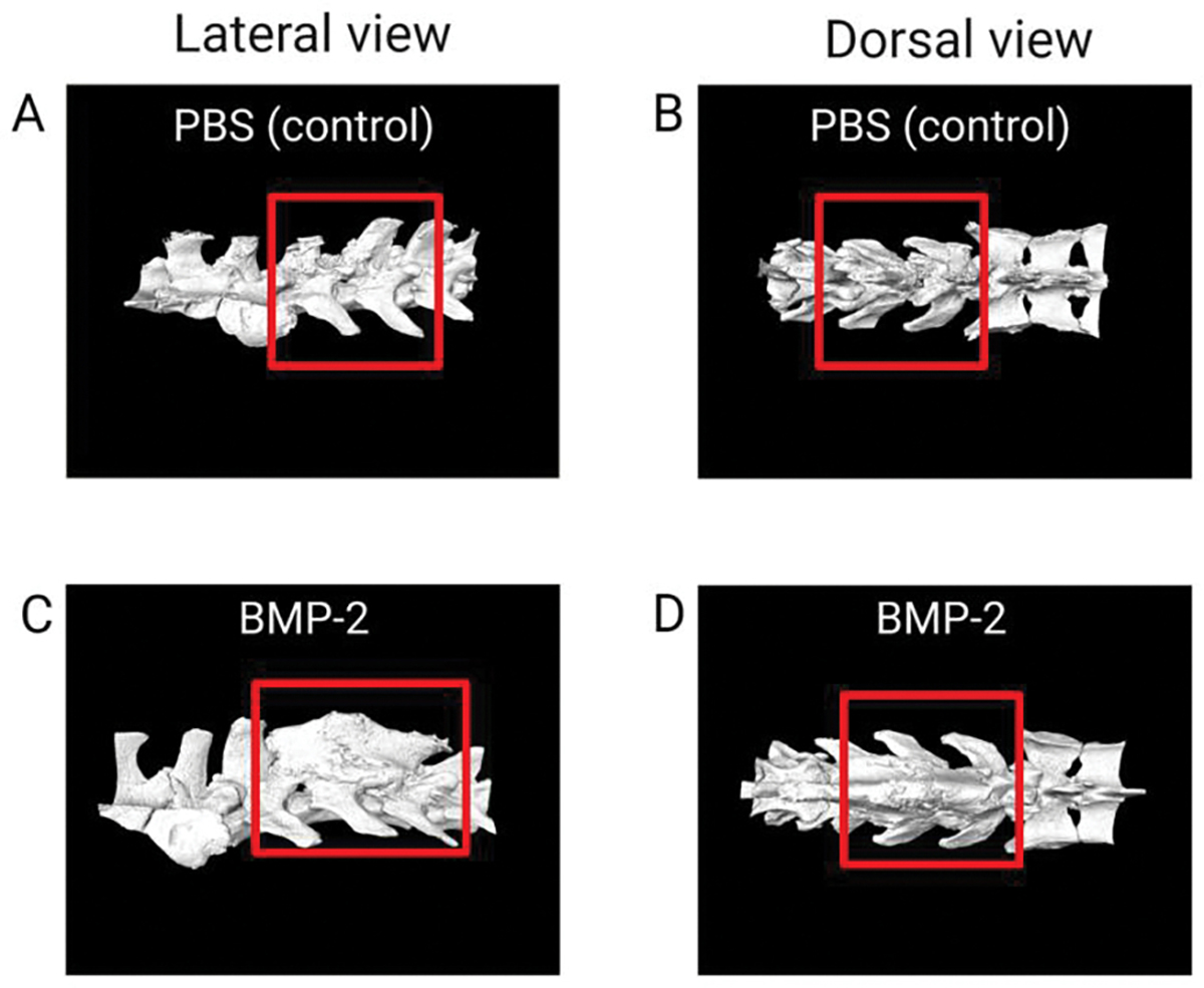
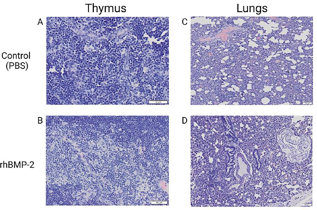
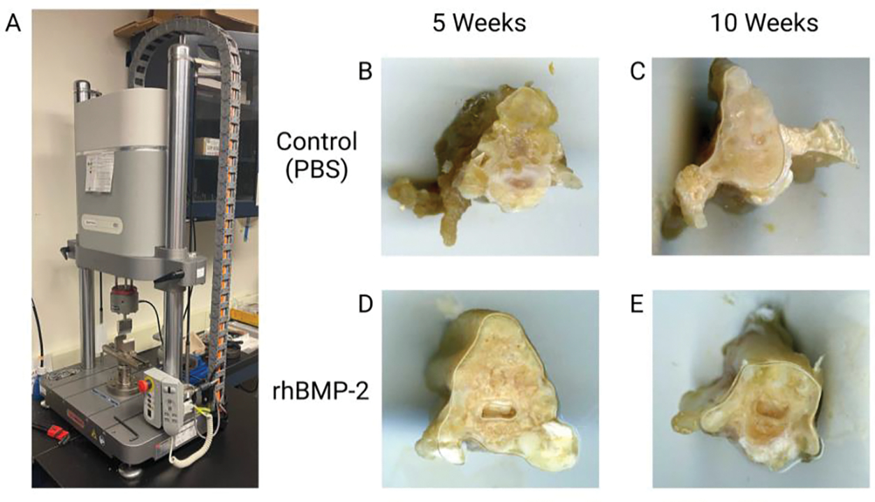
The rat animal model is a cost effective and reliable model used in spinal pre-clinical research. Complications from various surgical procedures in humans often arise that were based on these pre-clinical animal models. Therefore safe and efficacious pre-clinical animal models are needed to establish continuity into clinical trials. A Standard Operating Procedure (SOP) is a validated method that allows researchers to safely and carefully replicate previously successful surgical techniques. Thus, the aim of this study is to describe in detail the procedures involved in a common rat bilateral posterolateral intertransverse spinal fusion SOP used to test the efficacy and safety different orthobiologics using a collagen-soaked sponge as an orthobiologic carrier. Only two orthobiologics are currently FDA approved for spinal fusion surgery which include recombinant bone morphogenetic protein 2 (rhBMP-2), and I-FACTOR. While there are many additional orthobiologics currently being tested, one way to show their safety profile and gain FDA approval, is to use well established pre-clinical animal models. A preoperative, intraoperative, and postoperative surgical setup including specific anesthesia and euthanasia protocols are outlined. Furthermore, we describe different postoperative methods used to validate the spinal fusion SOP, which include μCT analysis, histopathology, biomechanical testing, and blood analysis. This SOP can help increase validity, transparency, efficacy, and reproducibly in future rat spinal fusion surgery procedures.

Keywords: Orthobiologics; Posterolateral intertransverse fusion model; Protocols; Rat model; Spinal fusion; Standard Operating Procedure (SOP); rhBMP-2.


Conflict of interest statement

Conflicts of Interest The authors have no conflicts of interest to declare.

Figures

Figure 1:
Figure 1:
Overview of the spinal fusion surgery steps. Outlined are the critical and major steps of the standard operating procedure developed from start to finish. The flow chart also includes the additional step of the caudectomy and morselization if the allograft is going to be used as a comparison.
Figure 2:
Figure 2:
Surgical instruments setup. Image A depicts the 1–4% isoflurane vaporizer setup with the nose cone lying in front of the metal tray where the animal will be placed. Image B depicts the Vetcorder animal vitals monitoring device. Image C depicts the surgical tray including forceps, scalpel, needle driver, suture scissors, curved forceps, and a retractor. Image D depicts the clear euthanasia chamber which is connected to 100% CO2.
Figure 3:
Figure 3:
Overview of procedure and workflow. Presented is a visual representation of the decortication of the spine and placement of the collagen sponge located at spinal levels L4-L5 in the rat. Additionally, the workflow of the post-operative procedure with the evaluation of μCT imaging, biomechanics, histology and blood analysis is shown.
Figure 4:
Figure 4:
Gross images of the spinal fusion during surgery and after dissection. Images in A depict different views of the decortication process and placement of the collagen sponge at the spinal fusion site. Images B and C, control (PBS) and rhBMP-2 respectively, are the dissected spines after animal sacrifice. The red boxes are approximately the L4-L5 area where the collagen sponge was placed.
Figure 5:
Figure 5:
μCT analysis of the spinal fusion site. Images A-D depicts the μCT images of the control in PBS and the treatment rhBMP-2. The red box outlines the L4-L5 area in which the spinal fusion bed was created, and collagen-soaked sponge was placed. Images A and C are the lateral views of the spine, with images B and D being the dorsal views of the spine. As shown in C and D in comparison to the control, rhBMP-2 increases bone regeneration at the level of the spinal fusion.
Figure 6:
Figure 6:
Thymus and lung samples of control and rhBMP-2 treatment. Images A-D depicts the histology staining and imaging post spinal fusion at 5 weeks. Images A and C show the control (PBS) while images B and D exhibit the rats treated with rhBMP-2. As presented in images B and D, rhBMP-2 induced a greater summation of inflammation (dark purple color) in samples as evidenced by increased quantity and morphed cell structure.
Figure 7:
Figure 7:
Biomechanical testing with 10mm sections of spinal fusion L4-L5. Image A is the Electropuls E3000 linear-torsion all-electric dynamic test instrument (Instron) used for biomechanical analysis of the spine sections. Image B and C are the controls (PBS) at 5 and 10 weeks respectively with image D and E being rhBMP-2 at 5 and 10 weeks, respectively.
Figure 8:
Figure 8:
Effects of estrogen on bone. Summarized chart of how estrogen affects different components of bone cells.

References

    1. Schnake KJ, Rappert D, Storzer B, et al. Lumbar fusion-Indications and techniques. Orthopade 48 (2019): 50–58. - PubMed
    1. Beckerman D, Esparza M, Lee SI, et al. Cost Analysis of Single-Level Lumbar Fusions. Global Spine J 10 (2020): 39–46. - PMC - PubMed
    1. Weiss AJ, Elixhauser A. Trends in Operating Room Procedures in U.S. Hospitals, 2001–2011: Statistical Brief #171, in Healthcare Cost and Utilization Project (HCUP) Statistical Briefs. Agency for Healthcare Research and Quality (US): Rockville (MD) (2006). - PubMed
    1. Dasta JF, McLaughlin TP, Mody SH, et al. Daily cost of an intensive care unit day: the contribution of mechanical ventilation. Crit Care Med 33 (2005): 1266–12 71. - PubMed
    1. White MS, Burns C, Conlon HA. The Impact of an Aging Population in the Workplace. Workplace Health Saf 66 (2018): 493–498. - PubMed

LinkOut - more resources