

Reconstruction of osseous defect with symphysis block graft for implant placement
- PMID: 36203858
- PMCID: PMC9531279
- DOI: 10.1016/j.jobcr.2022.09.010
Reconstruction of osseous defect with symphysis block graft for implant placement
Abstract
Introduction: Symphysis being an autogenous bone graft serves as one of the best graft for augmenting osseous defects of alveolar process with excellent results. It has been favoured mainly due to its local availability, accessibility and lesser resorption compared to other bones in the region.
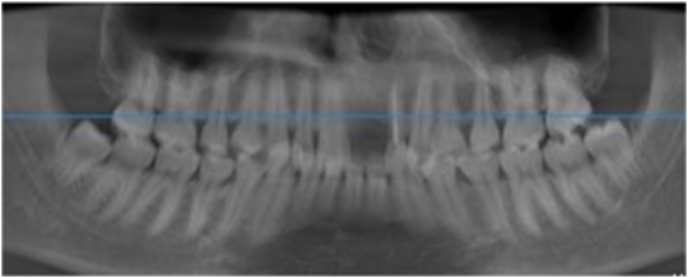
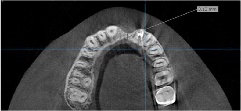
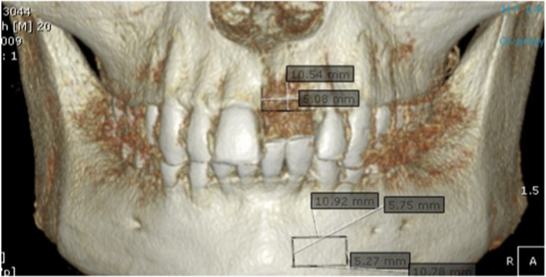
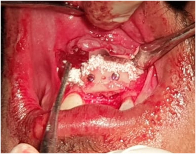
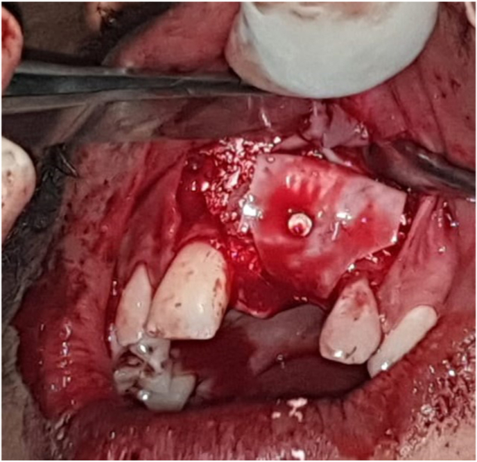
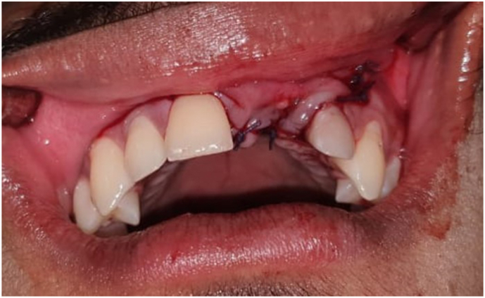
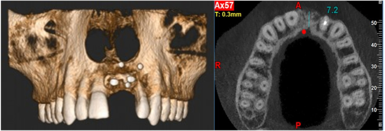
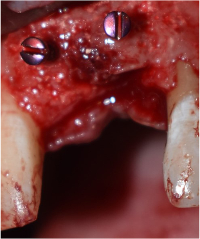
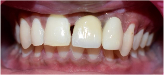
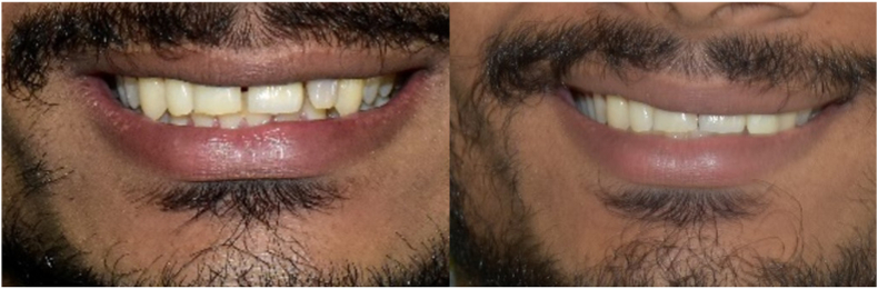
Case report: A 21/M reported to the department of Implantology with the complaint of missing tooth in the upper front tooth region since 1 year. History revealed extraction of upper left central incisor an year ago following trauma. Diagnosis was made as Siebert's Class I with horizontal bone loss irt 21 region with a bone defect of 10.54 x 5.08 x 4.85 mm. So a complete prosthetic rehabilitation protocol was made with an implant placement and grafting was planned with symphysis being most favourable.
Conclusion: The mandibular symphysis is a reliable intraoral graft site that can be used in the office setting with low morbidity. Because of the intraoral approach and lack of cutaneous scarring, patient acceptance is high.
Keywords: Autogenous graft; Implant grafting; Symphysis graft.
© 2022 Craniofacial Research Foundation. Published by Elsevier B.V.
Conflict of interest statement
None.
Figures


References
-
- Pikos M. Mandibular block Autografts for alveolar ridge augmentation. Atlas Oral Maxillofac Surg Clin. 2005;13(2):91–107. - PubMed
-
- Stern A., Barzani G. Dental Clinics of North America; 2015. Autogenous Bone Harvest for Implant Reconstruction. - PubMed
-
- Desai A., Mehta D., Tarun Kumar A., Thomas R. Current concepts and guidelines in chin graft harvesting: a literature review. Int J Occup Health Saf. 2013;3(1):16.
-
- Misch C.M., Misch C.E., Resnik R.R., Ismail Y.H. Reconstruction of maxillary alveolar defects with mandibular symphysis grafts for dental implants: a preliminary procedural report. Int J Oral Maxillofac Implants. 1992;7:360–366. - PubMed
-
- Pommer B., Tepper G., Gahleitner A., Zechner W., Watzek G. New safety margins for chin bone harvesting based on the course of the mandibular incisive canal in CT. Clin Oral Implants Res. 2008;19:1312–1316. - PubMed
LinkOut - more resources
Full Text Sources
