Review
. 2022 Jul 11;5(3):e1216.
doi: 10.1002/jsp2.1216. eCollection 2022 Sep.

Rat tail models for the assessment of injectable nucleus pulposus regeneration strategies

Affiliations
Review

Rat tail models for the assessment of injectable nucleus pulposus regeneration strategies

Marcos N Barcellona et al. JOR Spine. .

Abstract

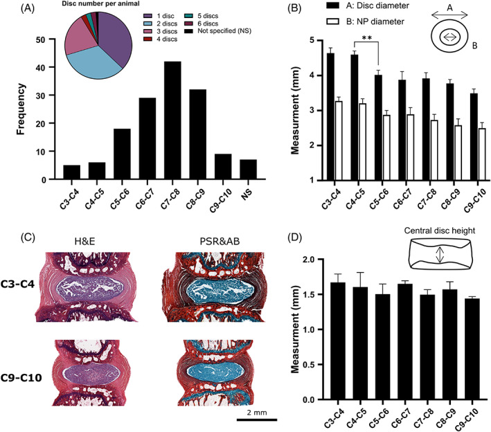
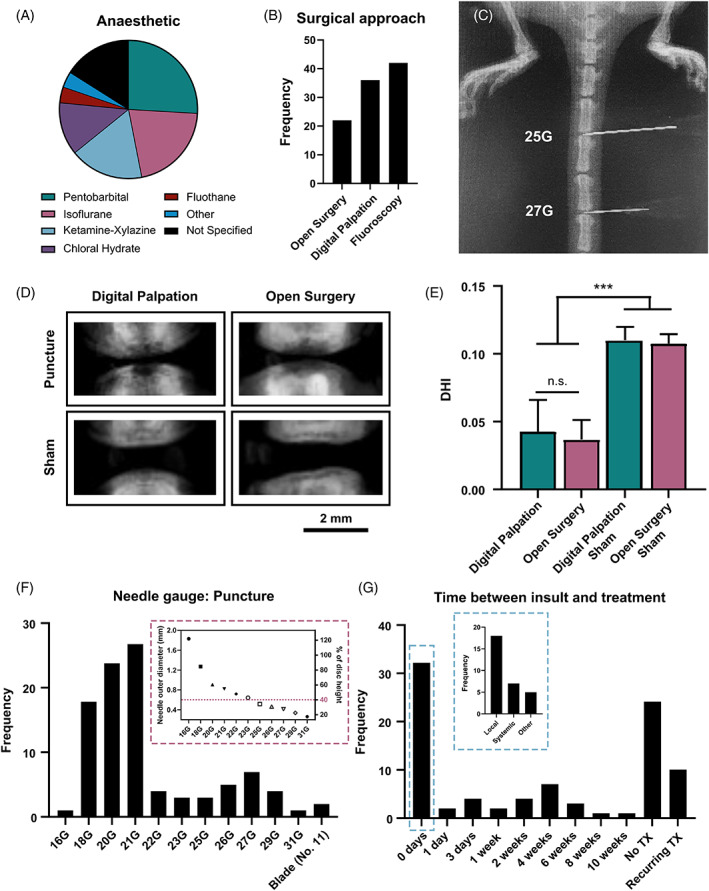
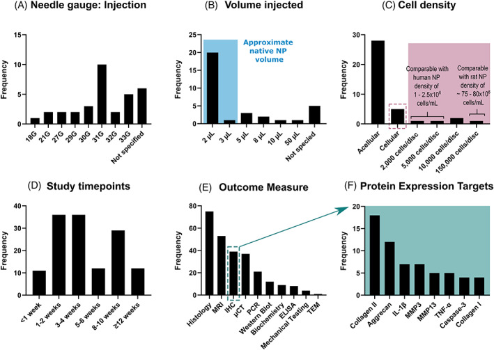
Back pain is a global epidemiological and socioeconomic problem often associated with intervertebral disc degeneration; a condition believed to initiate in the nucleus pulposus (NP). There is considerable interest in developing early therapeutic interventions to target the NP and halt degeneration. Rat caudal models of disc degeneration have demonstrated significant utility in the study of disease progression and its impact on tissue structure, composition, and mechanical performance. One significant advantage of the caudal model is the ease of access and high throughput nature. However, considerable variability exists across the literature in terms of experimental setup and parameters. The objective of this article is to aid researchers in the design and development of caudal puncture models by providing details and insight into the most reported experimental parameters. Preferred Reporting Items for Systematic Reviews and Meta-Analyses (PRISMA) guidelines were employed to screen the existing literature and 80 manuscripts met the inclusion criteria. Disc geometry, surgical approaches, effect of needle gauge size to induce degeneration, therapeutic volume, outcome measures, and associated limitations are considered and discussed, and a range of recommendations based on different research questions are presented.

Keywords: degeneration; intervertebral disc; nucleus pulposus; puncture; rat tail model; spine.


Conflict of interest statement

The authors declare no conflicts of interest.

Figures

FIGURE 1
FIGURE 1
Preferred Reporting Items for Systematic Reviews and Meta‐Analysis (PRISMA) diagram indicating the literature search terms, screening process, and exclusion criteria. One hundred and forty‐six nonduplicate manuscripts were identified across PubMed, Scopus, and Embase on March 7, 2022. Following screening and the application of exclusion criteria, 80 articles were included in this review
FIGURE 2
FIGURE 2
Summary of rat cohort, and the overall study design across the 80 articles reviewed. (A) The frequency of different rat strains used to create degeneration rat tail models (Sprague Dawley = 71.3%, Wister = 17.5%, Lewis = 5.0%, Athymic nude = 5.0% and 1.2% of studies did not specify rat strain). (B) Five studies (6.2%) used periadolescent rats at 6 weeks, while 26 studies (32.5%) used young adult rats at 8 and 10 weeks. However, most studies used rats older than 12 weeks, the timepoint at which skeletally maturity has occurred (12 weeks = 56.3%, 16 weeks = 1.3%, and 32 weeks = 2.5%). (C) Sixty‐five percent of studies used male rats, while only 12.0% of studies used female rats. However, 20.0% of the studies did not specify the sex of the animals used. (D) 41.3% of studies investigated a treatment delivered locally within the degenerated disc, 22.5% focused on systemic delivery such as incorporation of drugs into food, or intraperitoneal injections, while 30.0% solely developed a degeneration model with no treatment delivery
FIGURE 3
FIGURE 3
Anatomical and geometrical considerations. (A) The pie chart highlights the number of discs used per animal in each study, with most studies using 1 (37.5%), 2 (33.8%), or 3 (21.3%) discs. The bar graph shows the frequency and range of caudal levels used across these studies, with C7–C8 being the most frequent level and seven studies not specifying (NS) which discs were used. (B) Experimentally measured external disc diameter and internal nucleus pulposus (NP) diameter for 8‐week‐old Wister rats (N = 6). Statistics are only shown in the case of a statistical difference between adjacent levels with p = 0.0048. (C) Sagittal histology sections showing hematoxylin and eosin (H&E) and picrosirius red (PSR) combined with alcian blue (AB) for the most proximal and distal caudal discs investigated. (D) Experimentally measured central disc height, with no statistical significance found between adjacent levels within this range
FIGURE 4
FIGURE 4
Surgical approaches to establishing a degeneration rat tail model. (A) The majority of studies used pentobarbital (21/80 = 26.3%), isoflurane (17/80 = 21.3%), or ketamine–xylazine (14/80 = 17.5%). Less than 20% of studies used an alternative anesthetic; chloral hydrate, fluothane or other (medetomidine, midazolam, butorphanol, pelltobarbitalum natricum, or atipamezole). However, 13 studies (16.1%) did not report the anesthetic used. (B) The frequency of either digital palpation, open surgery, or fluoroscopically guided punctures used across the reviewed studies. (C) A microcomputed tomography (μCT) image demonstrating the fluoroscopically guided approach used to confirm the needle position. (D) μCT images to observe the reduction in disc height 2 weeks following subcutaneously puncture through digital palpation and open surgery. (E) Quantitative analysis using disc height index (DHI) to compare the effect of percutaneous puncture and open surgery (p < 0.003). (F) The frequency of needle gauges used to puncture the disc across the reviewed studies. The inset graph relates the outer needle diameter of each needle gauge to disc height, assuming an average disc height of 1.5 mm (G) The time between puncture insult and treatment (TX) across the reviewed studies
FIGURE 5
FIGURE 5
Treatment parameters and outcome measures used in the reviewed rat tail models. (A) Treatments are commonly administered through a 31G needle (10 papers). However, six papers failed to report on the needle gauge used. (B) Most studies reported an injection volume of 2 μl (20 studies), which is within the approximated rat NP volume estimated from NP tissue weights and accounting for hydration, blue overlay. (C) The majority of reviewed studies used an acellular approach (28 studies). Within the studies that used a cell‐based treatment (five studies) the cell densities varied extensively. Assuming a 2 μl NP volume, these cell densities have been compared to the native cell densities of the human and rat NP. (D) The literature reported a range of timepoints, with 1–2 weeks and 3–4 weeks the most popular for early timepoints and >8 weeks typical for later timepoints. (E) The frequency of different outcome measures used throughout the reviewed literature. (F) The most common protein expression targets determined through immunohistochemistry
FIGURE 6
FIGURE 6
A visual summary of the main findings compiled from the reviewed rat tail models. There is a general consensus to use rats at least 12 weeks old to ensure skeletal maturity, and both sexes are preferred. Most studies operate within the C5–C9 range. 18–21G needles are preferred for inducing degeneration, while a 31G needle is preferred for intradiscal treatment delivery. Pentobarbital and isoflurane are the favored anesthetics, followed by ketamine–xylazine and chloral hydrate. The surgical approach may be based on the specific experimental design and can include open surgery, fluoroscopy, and digital palpation. The most reported outcome measures were magnetic resonance imaging (MRI), microcomputed tomography (μCT) and histology. Created with BioRender.com

References

    1. Setton LA, Chen J. Mechanobiology of the intervertebral disc and relevance to disc degeneration. J Bone Joint Surg Am. 2006;88(2):52‐57. - PubMed
    1. Trout JJ, Buckwalter JA, Moore KC, Landas SK. Ultrastructure of the human intervertebral disc. I. Changes in notochordal cells with age. Tissue Cell. 1982;14(2):359‐369. - PubMed
    1. Shankar H, Scarlett JA, Abram SE. Anatomy and pathophysiology of intervertebral disc disease. Tech Reg Anesth Pain Manag. 2009;13(2):67‐75. doi:10.1053/J.TRAP.2009.05.001 - DOI
    1. Adams P, Eyre DR, Muir H. Biochemical aspects of development and ageing of human lumbar intervertebral discs. Rheumatology. 1977;16(1):22‐29. - PubMed
    1. Roberts S, Menage J, Urban JPG. Biochemical and structural properties of the cartilage end‐plate and its relation to the intervertebral disc. Spine (Phila pa 1976). 1989;14(2):166‐174. - PubMed