. 2022 Sep 23:22:1-17.
doi: 10.1016/j.bioactmat.2022.08.025. eCollection 2023 Apr.

Piezoresistive MXene/Silk fibroin nanocomposite hydrogel for accelerating bone regeneration by Re-establishing electrical microenvironment

Affiliations

Piezoresistive MXene/Silk fibroin nanocomposite hydrogel for accelerating bone regeneration by Re-establishing electrical microenvironment

Zhi-Chao Hu et al. Bioact Mater. .

Abstract

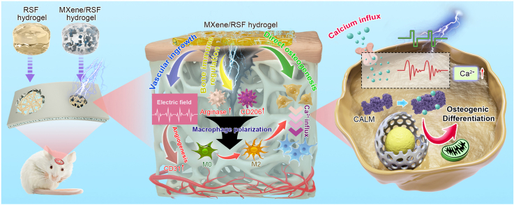
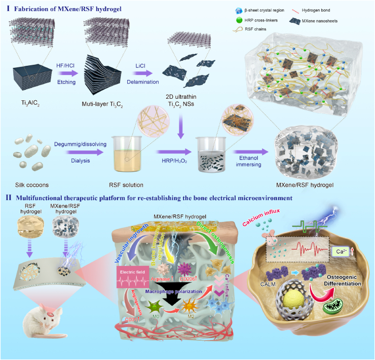
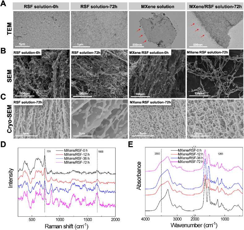
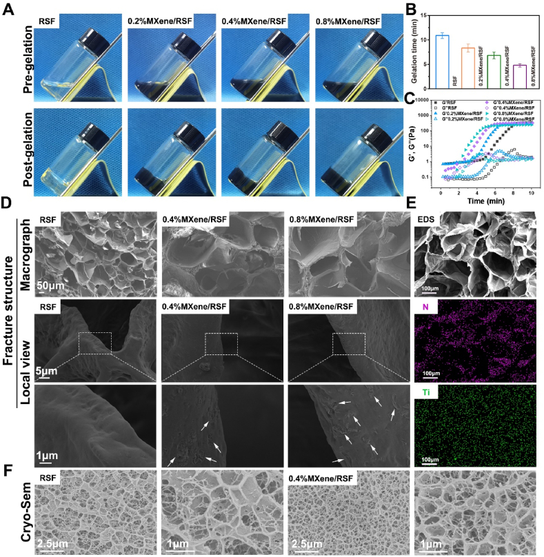
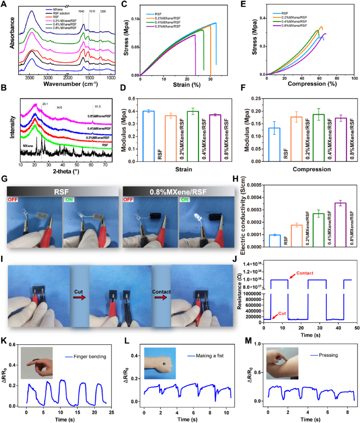
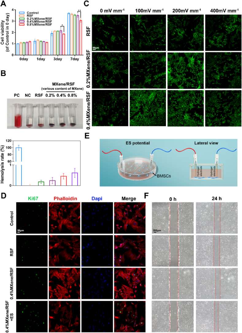
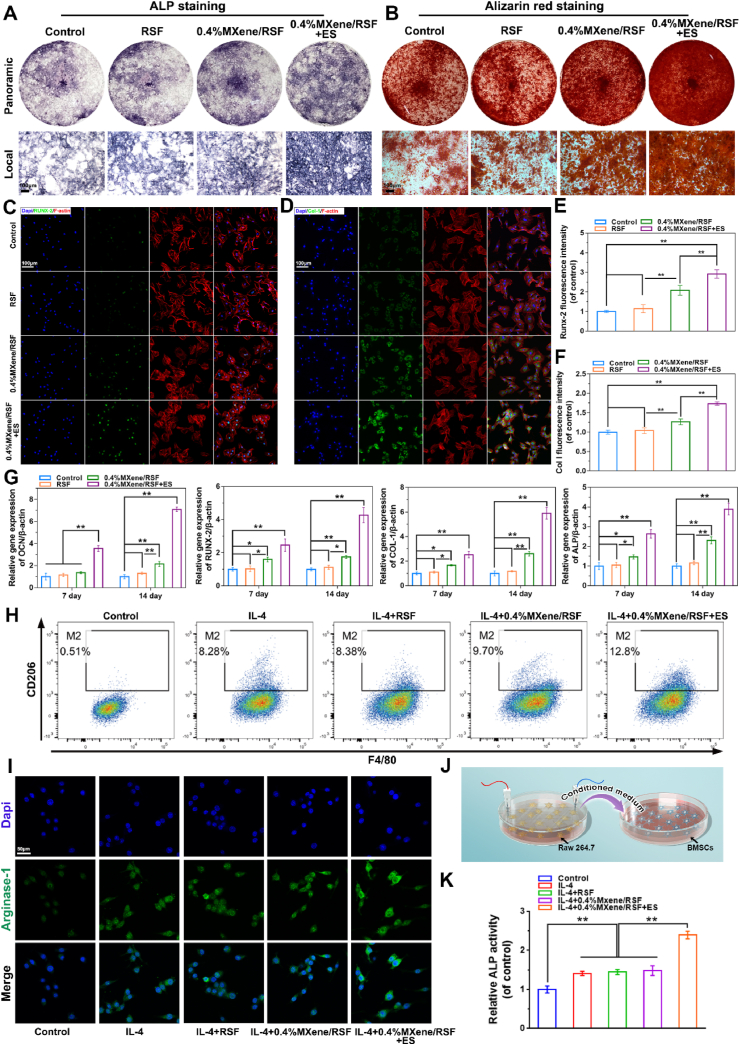
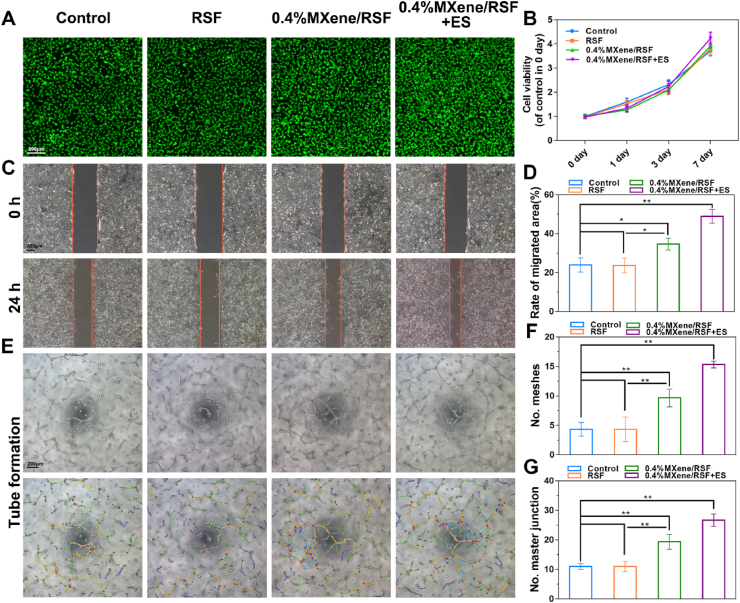
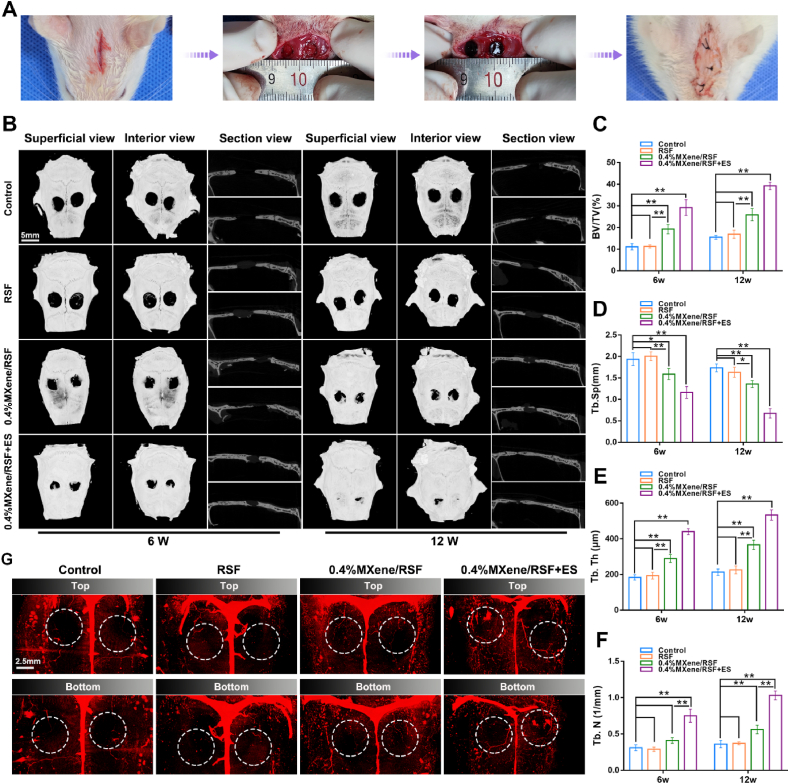
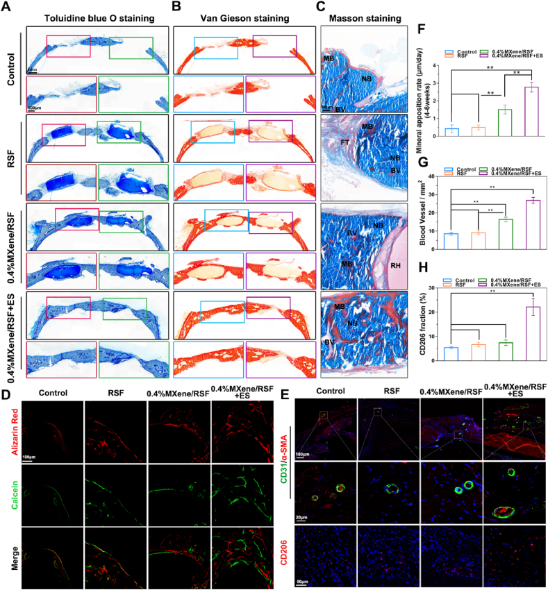
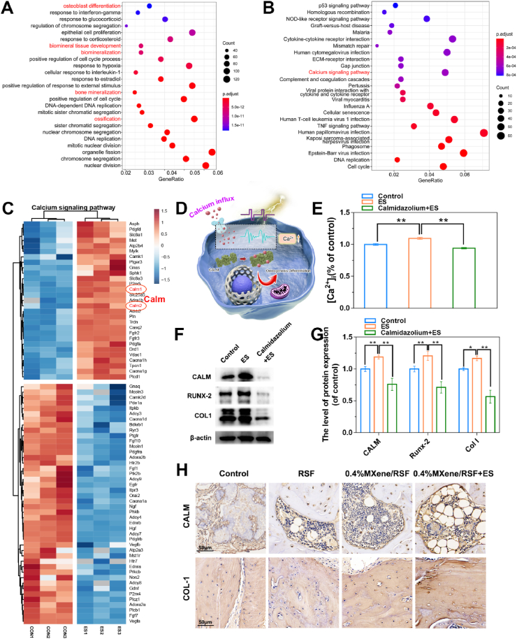
The electrical microenvironment plays an important role in bone repair. However, the underlying mechanism by which electrical stimulation (ES) promotes bone regeneration remains unclear, limiting the design of bone microenvironment-specific electroactive materials. Herein, by simple co-incubation in aqueous suspensions at physiological temperatures, biocompatible regenerated silk fibroin (RSF) is found to assemble into nanofibrils with a β-sheet structure on MXene nanosheets, which has been reported to inhibit the restacking and oxidation of MXene. An electroactive hydrogel based on RSF and bioencapsulated MXene is thus prepared to promote efficient bone regeneration. This MXene/RSF hydrogel also acts as a piezoresistive pressure transducer, which can potentially be utilized to monitor the electrophysiological microenvironment. RNA sequencing is performed to explore the underlying mechanisms, which can activate Ca2+/CALM signaling in favor of the direct osteogenesis process. ES is found to facilitate indirect osteogenesis by promoting the polarization of M2 macrophages, as well as stimulating the neogenesis and migration of endotheliocytes. Consistent improvements in bone regeneration and angiogenesis are observed with MXene/RSF hydrogels under ES in vivo. Collectively, the MXene/RSF hydrogel provides a distinctive and promising strategy for promoting direct osteogenesis, regulating immune microenvironment and neovascularization under ES, leading to re-establish electrical microenvironment for bone regeneration.

Keywords: Bone regeneration; Electrical microenvironment; Electrical stimulation; MXene; Regenerated silk fibroin.


Conflict of interest statement

The authors declare that they have no known competing financial interests or personal relationships that could have appeared to influence the work reported in this paper.

Figures

Image 1
Graphical abstract
Scheme 1
Scheme 1
Schematic showing the fabrication and application of multifunctional electroactive hydrogels for the diversified treatment of bone defects. (I) Fabrication process for an MXene/RSF hydrogel. (II) Conceptual and effective mechanisms of the electroactive MXene/RSF hydrogels to re-establish the electrical microenvironment for bone regeneration.
Fig. 1
Fig. 1
Characterization of the MXene-RSF interactions in solution. (A, B) TEM and SEM images of the pure RSF and MXene/RSF hybrid after assembly for 0 and 72 h. Red arrows indicate the morphology of the edge of MXene nanosheet. (C) Cryo-SEM images of the pure RSF and MXene/RSF hybrid after assembly for 72 h. (D) Raman spectra of MXene/RSF solution after incubation at 37 °C for different durations. (E) FT-IR spectra of MXene/RSF solution after incubation at 37 °C for different durations.
Fig. 2
Fig. 2
Gelation behavior and morphology characterization of the MXene/RSF hydrogels. (A) Digital photos of MXene/RSF solutions (up) and MXene/RSF hydrogels (down). (B) Gelation times for the different hydrogel samples with increasing concentrations of MXene. (C) Rheological properties exhibiting the G′ and G″ of various hydrogels. (D) SEM images showing the fracture morphology of hydrogel samples. (E) Fracture structure and element mapping of the MXene/RSF hydrogel. (F) Cryo-SEM images of the RSF and 0.4%MXene/RSF hydrogels.
Fig. 3
Fig. 3
Chemical structure and multifunctional properties of the MXene/RSF hydrogel. (A) FT-IR spectra of the RSF solution, MXene nanosheets, and RSF-based hydrogel samples. (B) XRD patterns of the different hydrogel samples. (C, D) Tensile stress–strain curves and elastic moduli for the different hydrogel samples. (E, F) Compressive stress–compression curves and compression moduli for the different hydrogel samples. (G) Electrically conductive pathways made of RSF and MXene/RSF hydrogels to illuminate an LED. (H) Electrical conductivity of hydrogels with various concentrations of MXene. (I, J) Cut-contact tests for the MXene/RSF hydrogel using a real-time resistance response. (K–M) Sensors assembled from MXene/RSF to monitor various human movements including finger bending, fisting, and pressing.
Fig. 4
Fig. 4
Proliferation and migration evaluation of the BMSCs with different hydrogels. (A) CCK-8 assay evaluating the cytotoxic effects after 1–7 days co-incubation with different hydrogels. (B) Hemolysis test for the RSF-based hydrogels. PC, a positive control (distilled water); NC, a negative control (normal saline). (C) Living/dead staining on different hydrogels with varying ES potentials. (D) Immunofluorescence staining results of Ki67 proteins. (E) A custom-designed device to detect cell migration using hydrogel electrodes. (F) Wound scratch assay in different hydrogel samples with or without ES potential. **P < 0.01, *P < 0.05, n = 3.
Fig. 5
Fig. 5
Osteogenic differentiation effects of the RSF-based hydrogel samples on the BMSCs and their immunomodulatory effect on macrophage polarization in RAW264.7 cells with or without ES. (A, B) Representative photographs of ALP staining on day 7 and ARS staining on day 21. (C–F) Representative immunofluorescence staining and quantitative analysis of Runx-2 and Col-1 proteins after 7 days. (G) Real-time PCR results for the mRNA expression of OCN, Runx-2, Col-1, and ALP on day 7 and 14. (H) Flow cytometry results of the percentage of CD206+F4/80+cells treated with different interventions for 48 h. (I) Representative immunofluorescence staining of Arginase-1. (J) Schematic diagram showing the process of osteogenic differentiation of BMSCs regulated by macrophage M2 polarization. (K) Quantitative analysis of ALP staining of BMSCs after being cultured using the conditioned medium of RAW264.7 cells for 7 days. **P < 0.01, *P < 0.05, n = 3.
Fig. 6
Fig. 6
Neovascularization stimulated by RSF-based hydrogels with or without ES. (A) Living/dead staining of HUVECs using different hydrogels with or without ES potential for 3 days. (B) Proliferation of HUVECs for 1, 3, and 7 days using the CCK-8 assay. (C, D) Wound scratch assay and quantitative analysis using HUVECs for 24 h. (E) Tube formation of HUVECs stimulated by RSF-based hydrogels and ES for 12 h. (F, G) Number of meshes and number of master junctions calculated using ImageJ. **P < 0.01, *P < 0.05, n = 3.
Fig. 7
Fig. 7
MXene/RSF hydrogels with ES potential accelerated bone regeneration. (A) Procedure for the establishment of the calvarial defect model in SD rats. (B) Cranial bone regeneration at 6 and 12 weeks after implantation presented by micro-CT. (C–F) BV/TV, Tb.Sp, Tb.Th, and Tb.N results calculated based on micro-CT. (G) Representative images of 3D reconstruction of the blood vessels at 12 weeks post-implantation. **P < 0.01, *P < 0.05, n = 3.
Fig. 8
Fig. 8
Histological staining and fluorescent imaging of the calvarial defect to assess new bone formation. (A) Toluidine blue, (B) van Gieson, and (C) Masson staining of undecalcified femoral sections at 12 weeks. IB, immature bone; MB, mature bone; BV, blood vessel; FT, fibrous tissue. (D) Representative images showing calcein and alizarin red staining. (E) Immunofluorescence staining for CD31 (green) and CD206 (red) in decalcified bone tissues. Nuclei were labeled using DAPI (blue). (F) Quantitative evaluation of MAR at 4–6 weeks post-implantation. (G) Quantitative analysis of the newly formed blood vessels at 12 weeks post-implantation. (H) Quantitative analysis of the CD206 positively stained cells. **P < 0.01, *P < 0.05, n = 3.
Fig. 9
Fig. 9
Underlying molecular mechanisms of osteogenesis induced by the ES-mediated MXene/RSF hydrogel. Top 25 results from the (A) GO and (B) KEGG biological process enrichment analysis of the upregulated and downregulated DEGs. (C) Heat map summarizing the DEGs related to the calcium-signaling pathway. (D) Schematic diagram showing the potential calcium-signaling pathway. (E) Relative intracellular calcium ion concentration ([Ca2+]i) of BMSCs on day 7. (F, G) Protein expression levels and quantitative results of CALM, RUNX-2, and COL-1 in BMSCs with various interventions as indicated. (H) Immunohistochemical staining of CALM and COL-1 in decalcified bone tissue at 12 weeks after surgery. **P < 0.01, *P < 0.05, n = 3.

References

    1. Catterall W.A., Wisedchaisri G., Zheng N. The chemical basis for electrical signaling. Nat. Chem. Biol. 2017;13(5):455–463. - PMC - PubMed
    1. Rajabi A.H., Jaffe M., Arinzeh T.L. Piezoelectric materials for tissue regeneration: a review. Acta Biomater. 2015;24:12–23. - PubMed
    1. Thrivikraman G., Lee P.S., Hess R., Haenchen V., Basu B., Scharnweber D. Interplay of substrate conductivity, cellular microenvironment, and pulsatile electrical stimulation toward osteogenesis of human mesenchymal stem cells in vitro. ACS Appl. Mater. Interfaces. 2015;7(41):23015–23028. - PubMed
    1. Mollon B., da Silva V., Busse J.W., Einhorn T.A., Bhandari M. Electrical stimulation for long-bone fracture-healing: a meta-analysis of randomized controlled trials. J. Bone Joint Surg. Am. 2008;90(11):2322–2330. - PubMed
    1. Eischen-Loges M., Oliveira K.M.C., Bhavsar M.B., Barker J.H., Leppik L. Pretreating mesenchymal stem cells with electrical stimulation causes sustained long-lasting pro-osteogenic effects. PeerJ. 2018;6:e4959. - PMC - PubMed

LinkOut - more resources