. 2022 Sep 12:2022:9827986.
doi: 10.34133/2022/9827986. eCollection 2022.

Molecular Mechanisms of Spawning Habits for the Adaptive Radiation of Endemic East Asian Cyprinid Fishes

Affiliations

Molecular Mechanisms of Spawning Habits for the Adaptive Radiation of Endemic East Asian Cyprinid Fishes

Feng Chen et al. Research (Wash D C). .

Abstract

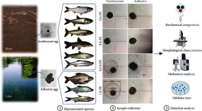
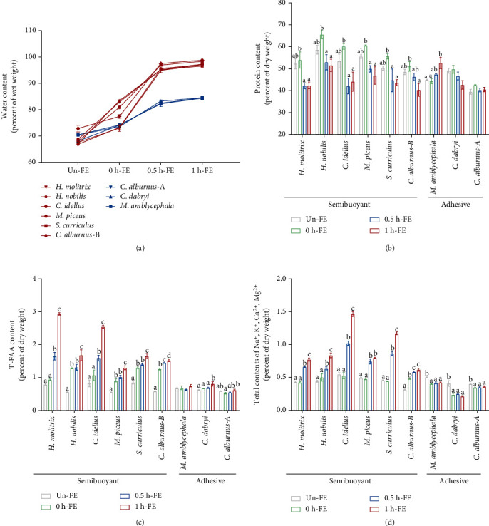
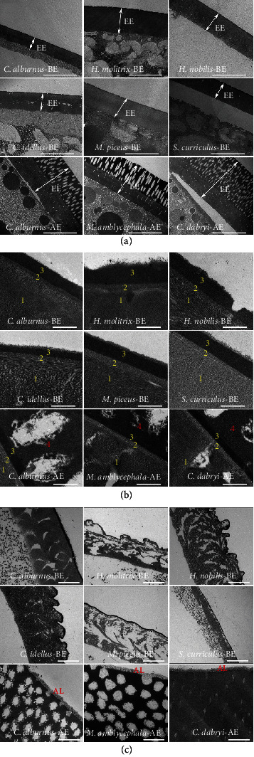
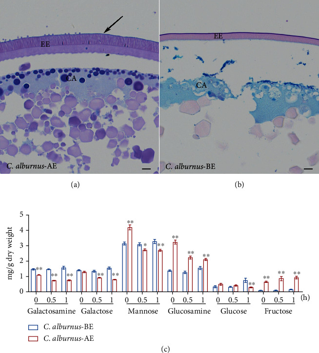
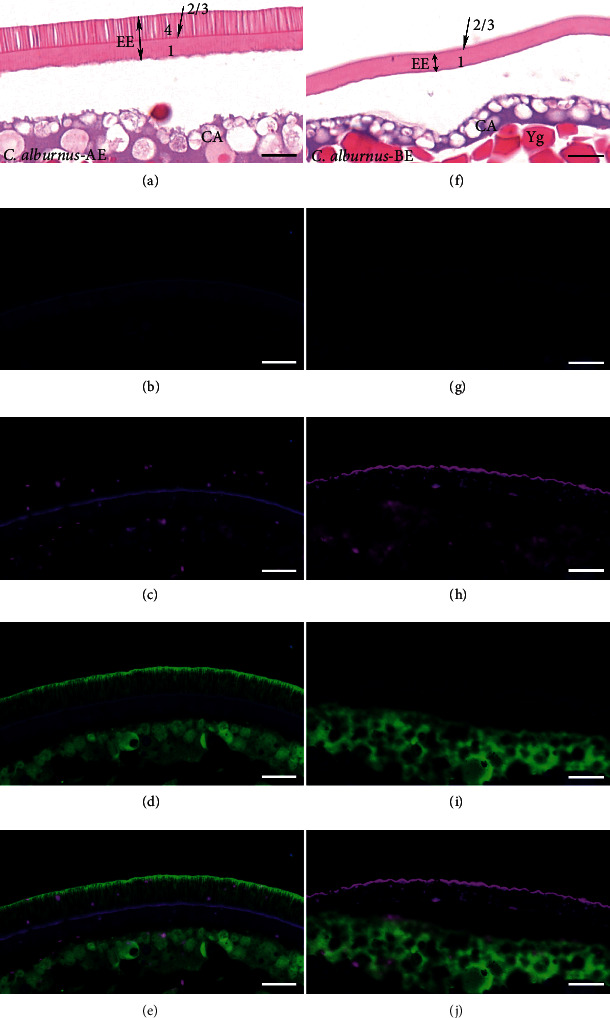
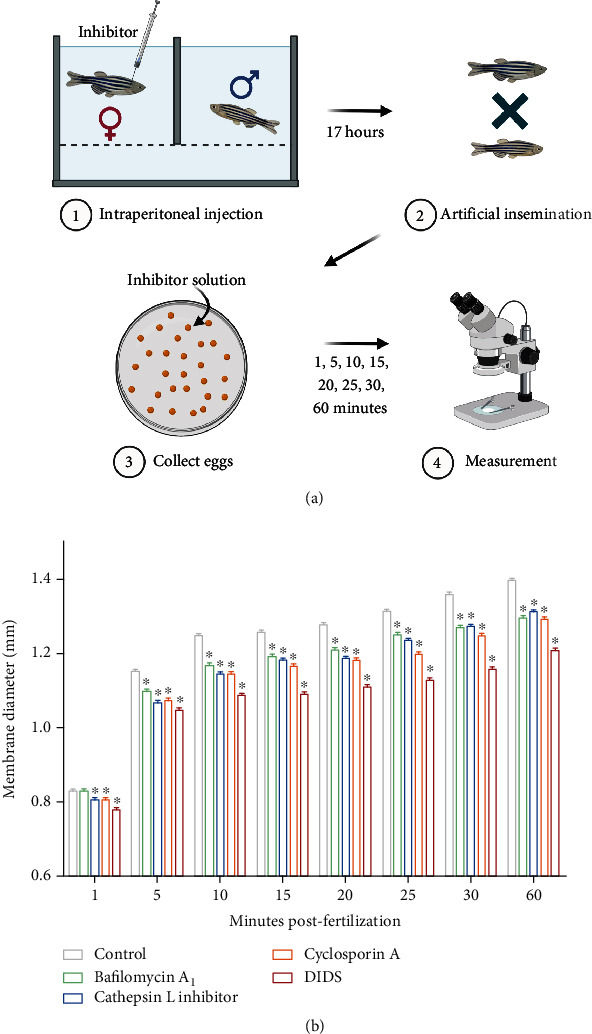
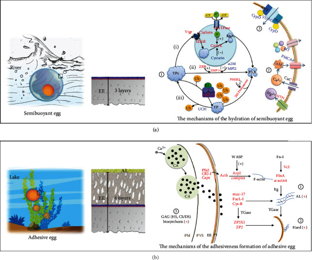
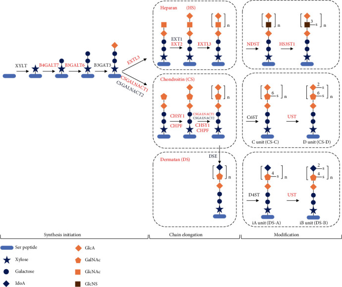
Despite the widespread recognition of adaptive radiation as a driver of speciation, the mechanisms by which natural selection generates new species are incompletely understood. The evolutionary radiation of endemic East Asian cyprinids has been proposed as evolving through a change in spawning habits, involving a transition from semibuoyant eggs to adhesive eggs in response to crosslinked river-lake system formation. Here, we investigated the molecular mechanisms that underpin this radiation, associated with egg hydration and adhesiveness. We demonstrated that semibuoyant eggs enhance hydration by increasing the degradation of yolk protein and accumulation of Ca2+ and Mg2+ ions, while adhesive eggs improve adhesiveness and hardness of the egg envelope by producing an adhesive layer and a unique 4th layer to the egg envelope. Based on multiomics analyses and verification tests, we showed that during the process of adaptive radiation, adhesive eggs downregulated the "vitellogenin degradation pathway," "zinc metalloprotease pathway," and "ubiquitin-proteasome pathway" and the pathways of Ca2+ and Mg2+ active transport to reduce their hydration. At the same time, adhesive eggs upregulated the crosslinks of microfilament-associated proteins and adhesive-related proteins, the hardening-related proteins of the egg envelope, and the biosynthesis of glycosaminoglycan in the ovary to generate adhesiveness. These findings illustrate the novel molecular mechanisms associated with hydration and adhesiveness of freshwater fish eggs and identify critical molecular mechanisms involved in the adaptive radiation of endemic East Asian cyprinids. We propose that these key egg attributes may function as "magic traits" in this adaptive radiation.


Conflict of interest statement

The authors declare that there is no conflict of interest regarding the publication of this article.

Figures

Figure 1
Figure 1
A schematic of the habitats associated with semibuoyant and adhesive eggs and the experimental design and data collection. Six representative species (H. molitrix, H. nobilis, C. idella, M. piceus, S. curriculus, and C. alburnus-B) of the endemic East Asian cyprinid group that produces semibuoyant eggs in lotic (rivers) habitats and three representative species (M. amblycephala, C. dabryi, and C. alburnus-A) of the endemic group that spawn adhesive eggs in lentic (lakes) habitats. Semibuoyant eggs and adhesive eggs were collected unfertilized (Un-FE) and 0, 0.5, and 1 h post fertilization (0 h-FE, 0.5 h-FE, and 1 h-FE) and used for elucidating the mechanisms of hydration and adhesiveness. MD: membrane diameter; ED: egg diameter.
Figure 2
Figure 2
Contrasting spawning habits differ in their biochemical composition of unfertilized eggs (Un-FE) and 0, 0.5, and 1 h post fertilization (0 h-FE, 0.5 h-FE, and 1 h-FE). (a) The water contents of semibuoyant eggs (red lines) and adhesive eggs (blue lines). (b) The protein contents, (c) T-FAA contents, and (d) the total contents of Na+, K+, Ca2+, and Mg2+ ions of different spawning habits. Values are presented as the mean ± SEM from three to five separate experiments. Different lowercase letters indicate significant differences among the four time periods (p < 0.05, one-way analysis of variance). T-FAA: total free amino acid.
Figure 3
Figure 3
The egg envelope ultrastructure of six representative species with semibuoyant eggs (C. alburnus-B, H. molitrix, H. nobilis, C. idella, M. piceus, and S. curriculus) and three representative species with adhesive eggs (C. alburnus-A, M. amblycephala, and C. dabryi) of endemic East Asian cyprinids. (a) TEM images of the egg envelopes of unfertilized eggs (scale bar is 10 μm). EE: egg envelope. (b) TEM images of the egg envelopes of unfertilized eggs (scale bar is 500 nm). The egg envelopes of semibuoyant eggs were divided into three layers (1, 2, and 3), and the egg envelopes of adhesive eggs were divided into four layers (1, 2, 3, and 4). (c) TEM images of the adhesive layer of eggs at 1 h post fertilization (scale bar is 2 μm). AL: adhesive layer; BE: semibuoyant egg; AE: adhesive egg.
Figure 4
Figure 4
Histological characterization and monosaccharides of the egg envelope in two spawning habits of C. alburnus eggs after fertilization. (a, b) Histological characterization staining with Alcian blue- (pH 2.5) periodic acid Schiff (AB-PAS) in the adhesive and semibuoyant eggs of C. alburnus at 1 h after fertilization (scale bar is 10 μm). Black arrow: outer edge of the 4th layer of the adhesive egg envelope; AE: adhesive egg; BE: semibuoyant egg; EE: egg envelope; CA: cortical alveoli. (c) The monosaccharide analysis of the egg envelopes of the two types of C. alburnus eggs was performed at 0, 0.5, and 1 h after fertilization. Values are presented as the mean ± SEM. indicates p < 0.05 versus C. alburnus-BE and ∗∗indicates p < 0.01 versus C. alburnus-BE.
Figure 5
Figure 5
Co-immunofluorescence analysis demonstrating localization of egg envelope proteins (ZP2 and ZP3X1) in unfertilized eggs (scale bar is 20 μm). (a–e) Adhesive egg of C. alburnus (C. alburnus-AE); (f–j) semibuoyant egg of C. alburnus (C. alburnus-BE); (a, f) stained results with haematoxylin-eosin; (b, g) control groups; (c–e and h–j) immunofluorescence localization of ZP2 (magenta) and ZP3X1 (green) in the egg envelope. EE: egg envelope; CA: cortical alveoli; Yg: yolk globule.
Figure 6
Figure 6
A scheme for the experimental design and the membrane diameter of zebrafish eggs after inhibition. (a) Mature female zebrafish were treated with 0.05% DMSO, 100 nM bafilomycin A1, 1.5 μM cathepsin L inhibitor, 5 μM cyclosporine A, and 200 μM DIDS by intraperitoneal injection (n = 3). After artificial insemination, zebrafish eggs were immersed in DMSO or inhibitor solutions and their membrane diameter was measured at 1, 5, 10, 15, 20, 25, 30, and 60 minutes post fertilization. (b) The membrane diameter of zebrafish eggs at 1, 5, 10, 15, 20, 25, 30, and 60 minutes post fertilization when exposed to DMSO or inhibitor solutions. Values are presented as the mean ± SEM from three separate experiments (30–40 per group in each experiment). Asterisks indicate significant differences between the control and treatment groups (p < 0.05, Student's t-test). DMSO: dimethyl sulfoxide; DIDS: 4,4′-diisothiocyanatostilbene-2,2′-disulfonic acid disodium salt.
Figure 7
Figure 7
A schematic summary of the molecular mechanisms of two spawning habits for the adaptive radiation of endemic East Asian cyprinids. (a) The mechanisms underlying the hydration of semibuoyant eggs. ① Three pathways of yolk protein degradation. (i) Vtg degradation pathway, (ii) zinc metalloproteinase pathway, and (iii) ubiquitin-proteasome pathway. ② The pathways of Ca2+ and Mg2+ active transport. ③ The molecular structure of the egg envelope permeability transition pore. Proteins in red and blue show significant upregulation and downregulation in semibuoyant eggs, respectively. (b) The mechanisms underlying adhesiveness formation of adhesive eggs. ① The crosslinks of microfilament-associated proteins and adhesive-related proteins. ② The hardening of the egg envelope. Proteins in red show significant upregulation in adhesive eggs. Details are shown in Tables S2 and S4. ③ The biosynthesis of glycosaminoglycan (GAG) in the ovary, including the biosynthesis of immediate precursors of GAG determined by proteomics analysis (details are shown in Table S3 and Figure S8) and the synthesis and modification of heparan sulphate (HS) and chondroitin/dermatan sulphates (CS/DS) by transcriptomics analysis (details are shown in Table S6 and Figure 8). EE: egg envelope; PVS: perivitelline space; Yp: yolk protein; FAA: free amino acids; Vtg: vitellogenin; Ub: ubiquitin; CaC: Ca2+ channel; CA: cortical alveoli; PM: plasma membrane; AL: adhesive layer.
Figure 8
Figure 8
The transcription level of biosynthesis of glycosaminoglycan (GAG) in the ovary, including heparan sulphate (HS) and chondroitin/dermatan sulphate (CS/DS) core structures and modification. Genes in red involved in synthesis initiation, chain elongation, and modification in the biosynthesis of GAG are significantly upregulated in ovaries producing adhesive eggs. Details are shown in Table S6. Ser peptide: light blue ovals; xylose residue: blue star; galactose: blue circle; iduronic acid (IdoA): blue diamond; glucuronic acid (GlcA): orange diamond; N-acetylgalactosamine (GalNAc): orange pentagon; N-acetylglucosamine (GlcNAc): orange square; N-sulfoglucosamine (GlcNS): brown square. B4GALT7: β-1,4-galactosyltransferase 7; B3GALT6: β-1,3-galactosyltransferase 6; EXTL3: exostosin-like 3; EXT1: exostosin-1b; EXT2: exostosin-2; NDST: heparan sulphate N-deacetylase/N-sulfotransferase; HS3ST1: heparan sulphate glucosamine 3-O-sulfotransferase 1; CSGALNACT1: chondroitin sulphate N-acetylgalactosaminyltransferase 1; CSGALNACT2: chondroitin sulphate N-acetylgalactosaminyltransferase 2; CHSY1: chondroitin sulphate synthase 1; CHPF: chondroitin polymerizing factor a; UST: uronyl 2-sulfotransferase; DSE: dermatan-sulphate epimerase; D4ST: dermatan 4 sulfotransferase 1.

References

    1. Darwin C. On the Origin of Species . John Murray; 1859.
    1. Marques D. A., Meier J. I., Seehausen O. A combinatorial view on speciation and adaptive radiation. Trends in Ecology & Evolution . 2019;34(6):531–544. doi: 10.1016/j.tree.2019.02.008. - DOI - PubMed
    1. McGee M. D., Borstein S. R., Meier J. I., et al. The ecological and genomic basis of explosive adaptive radiation. Nature . 2020;586(7827):75–79. doi: 10.1038/s41586-020-2652-7. - DOI - PubMed
    1. Schluter D. The Ecology of Adaptive Radiation . Oxford: OUP; 2000.
    1. Zhang Y., Teng D., Lu W., et al. A widely diverged locus involved in locomotor adaptation in Heliconius butterflies. Science Advances . 2021;7(32, article eabh2340) doi: 10.1126/sciadv.abh2340. - DOI - PMC - PubMed

LinkOut - more resources