

Case report: Spontaneous closure of ventricular pseudoaneurysm post-acute myocardial infarction with non-surgical therapy
- PMID: 36204582
- PMCID: PMC9530630
- DOI: 10.3389/fcvm.2022.996072
Case report: Spontaneous closure of ventricular pseudoaneurysm post-acute myocardial infarction with non-surgical therapy
Abstract
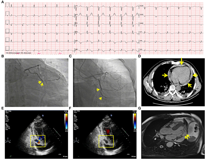
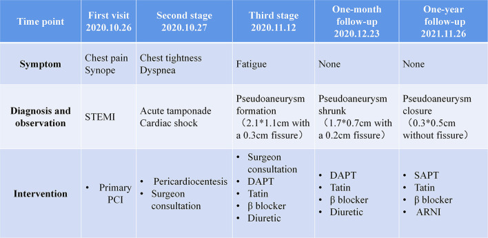
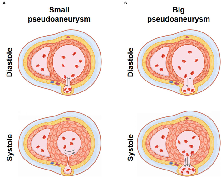
Left ventricle (LV) pseudoaneurysm is a rare disorder post-acute myocardial infarction (AMI). Resection or closure of the pseudoaneurysm by surgery is recommended due to the high propensity of pseudoaneurysm rupture while surgery has also high risks. Conservative therapy could be acceptable in small pseudoaneurysms or patients with high surgical risks. Nevertheless, the risk evaluation and grasp of indication are not clear. This case reported an acute cyst-like LV pseudoaneurysm formation post-AMI-induced myocardial free wall rupture (MFWR), and the patient recovered with spontaneous closure of the fissure and shrinkage of the LV pseudoaneurysm through non-surgical therapy. Based on the observations in the echocardiogram, we proposed that intermittent closing of the fissure and interruption of the blood flow between the LV and the pseudoaneurysm due to LV contraction alleviated stress change on the pseudoaneurysm. The narrow fissure, small pseudoaneurysm, and intermittently interrupted blood flow that benefit fissure healing and pseudoaneurysm stabilization could indicate the prognosis of this patient. Drugs like β-blocker that decreased the stress on the pseudoaneurysm also led to the risk reduction of pseudoaneurysm rupture. To our knowledge, this is the first case that reports a spontaneous closure of LV pseudoaneurysm. The size of the fissure and the pseudoaneurysm, as well as the corresponding hemodynamic state, could be valuable to evaluate the risk and prognosis of the pseudoaneurysm. Optimized medical management was also helpful to pseudoaneurysm stabilization.
Keywords: conservative therapy; hemodynamic; myocardial infarction; outcomes; shape; ventricular pseudoaneurysm.
Copyright © 2022 Shuai, Hu and Wei.
Conflict of interest statement
The authors declare that the research was conducted in the absence of any commercial or financial relationships that could be construed as a potential conflict of interest.
Figures
References
-
- Frances C, Romero A, Grady D. Left ventricular pseudoaneurysm. J Am Coll Cardiol. (1998) 32:557–61. - PubMed
Publication types
LinkOut - more resources
Full Text Sources