Review
. 2023 Apr;18(4):701-707.
doi: 10.4103/1673-5374.354511.

Neural and Müller glial adaptation of the retina to photoreceptor degeneration

Affiliations
Review

Neural and Müller glial adaptation of the retina to photoreceptor degeneration

Henri O Leinonen et al. Neural Regen Res. 2023 Apr.

Abstract

The majority of inherited retinal degenerative diseases and dry age-related macular degeneration are characterized by decay of the outer retina and photoreceptors, which leads to progressive loss of vision. The inner retina, including second- and third-order retinal neurons, also shows aberrant structural changes at all stages of degeneration. Müller glia, the major glial cells maintain retinal homeostasis, activating and rearranging immediately in response to photoreceptor stress. These phenomena are collectively known as retinal remodeling and are anatomically well described, but their impact on visual function is less well characterized. Retinal remodeling has traditionally been considered a detrimental chain of events that decreases visual function. However, emerging evidence from functional assays suggests that remodeling could also be a part of a survival mechanism wherein the inner retina responds plastically to outer retinal degeneration. The visual system´s first synapses between the photoreceptors and bipolar cells undergo rewiring and functionally compensate to maintain normal signal output to the brain. Distinct classes of retinal ganglion cells remain even after the massive loss of photoreceptors. Müller glia possess the regenerative potential for retinal recovery and possibly exert adaptive transcriptional changes in response to neuronal loss. These types of homeostatic changes could potentially explain the well-maintained visual function observed in patients with inherited retinal degenerative diseases who display prominent anatomic retinal pathology. This review will focus on our current understanding of retinal neuronal and Müller glial adaptation for the potential preservation of retinal activity during photoreceptor degeneration. Targeting retinal self-compensatory responses could help generate universal strategies to delay sensory disease progression.

Keywords: Müller glia; bipolar cells; electroretinography; photoreceptors; plasticity; retinal degeneration; retinal ganglion cells; retinal neuron; retinal remodeling.


Conflict of interest statement

None

Figures
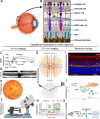
Figure 1
Figure 1
Schematics of retinal structure and established technologies for retina research. (A) The transparency of the eye allows direct visual access to the retina, which is the only neural tissue that is exposed to visual inspection noninvasively. (B) The layered and strictly hierarchical architecture of the retina is arguably the best characterized neural system in our bodies. (C) Electroretinogram (ERG) is a minimally invasive method to inspect sensory neuron (photoreceptor) activation (a-wave) and synaptic transmission to interneurons (b-wave). (D) Optical coherence tomography (OCT) allows segmentation of the retina noninvasively. (E) Scanning laser ophthalmoscopy (SLO) allows high-resolution images of vasculature. (F) Several advanced retina imaging methods using fluorescence, including 2-photon excitation, are established. (G) Dissected and perfused live retina is highly amenable to electrophysiology by e.g. multielectrode arrays (MEA) and patch-clamp, and (H) to assessment of metabolism. (I) Retina’s distinctly layered anatomy renders histological inspection and immunohistochemistry very practical. RGCL: Retinal ganglion cell layer; IPL: inner plexiform layer; INL: inner nuclear layer; OPL: outer plexiform layer; ONL: outer nuclear layer. (J) Single-cell transcriptomics maps gene profile at specific cells. (K) Established methods exist for preparation of primary cell cultures of several main cell classes in the retina. Graphs A, B, E, F, G, H, and K were adapted/reprinted from “Eye and Retina schematics”, “Diabetic Retinopathy Hallmarks”, “Rodent Fundoscopy”, “Retina MEA”, “Retinal ganglion cell patch clamp”, “Metabolic Assays -Using Seahorse Analyzers” and “Primary Cell Culture Preparation” with BioRender.com (2022). Retrieved from https://app.biorender.com/biorender-templates (license #: ZR241JTGY4, BU241JTVMS, NW241JU576, AQ241JUC41, LR241JUJWJ, JB241JVDIA, RD241JUX5O). Graphs C, D, I, and J present unpublished data from the Leinonen Retina Laboratory.
Figure 2
Figure 2
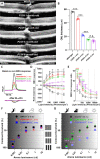
Near normal rod-dependent visual contrast sensitivity remains for several months in retinitis pigmentosa mice. (A) Representative optical coherence tomography (OCT) images showing degeneration progression in P23H mice. (B) Mean outer nuclear layer (ONL, photoreceptor nuclei layer) thickness as measured at 500 µm from the optic nerve head at dorsal, ventral, nasal, and temporal retinal quadrants. (C) Representative ex vivo ERG responses for dim flash (17 photons/µm2) in GNAT2–/– control and GNAT2–/–/P23H retinitis pigmentosa mice at baseline and after DL-AP4 perfusion to isolate rod-specific response. (D) Rod bipolar cells (RBC) and rod response amplitudes. RBC response was acquired offline by digitally subtracting the rod-specific response from the basal response. (E) RBC/rod response ratio. (F, G) Visual contrast thresholds as measured by optomotor responses (OMR) in 2–3-month-old (F) and 5–6-month-old (G) mice. Note the abrupt drop in ERGs and OMR performance between 5 and 6 months of age in GNAT2–/–/P23H mice. Adapted from Leinonen et al. (2020).
Figure 3
Figure 3
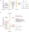
Müller glial responses in retinal degeneration. (A) Müller glial regeneration in zebrafish. Upon injury, Müller glia undergo reprogramming and enter the cell cycle for proliferation, stimulated by β-catenin, Mapk/Erk, Jak/signal transducer and activator of transcription 3 (Stat3), PI3K/Akt. These signaling pathways further induce Ascl expression to generate Müller glial progenitor cells, which regenerate the injured retina. Growth factors and cytokines (such as FGF2, HBEGF, insulin, Midkine-a, TNFα) as well as Wnts positively stimulate the process. TGF-β, Notch, and neurotransmitter (GABA, glutamate) signaling negatively regulates the process. Adapted and updated from Salman et al. (2021). (B) In mammalian retina, the regeneration potential of Müller glia is limited. The activated Müller glial may undergo metabolic adaptation and remodeling to preserve neural retinal function. Interestingly, EGF and HBEGF stimulate while TGF-β suppresses Müller glial proliferation in mammalian retinas. Manipulation of β-catenin, Nfia/b/x, Hippo, and YAP pathways stimulates the generation of Müller glial progenitor cells, which can be further differentiated into photoreceptors upon the subsequent activation of Otx2, Crx, and Nrl following the stimulation of the Wnt pathway by targeting β-catenin. The graph was adapted/reprinted from “retinal cell (Müller glia)”, “cone photoreceptor”, “rod photoreceptor”, “retinal cell (ganglion 1)”, “bipolar neuron”, “retinal cell (amacrine)”, “retinal cell (horizontal)” by BioRender.com (2022). Retrieved from https://app.biorender.com/biorender-templates (license #:HT240EKBMT). Ascl: Achaete-scute complex-like 1; Crx: cone-rod homeobox; EGF: epidermal-like growth factor; FGF2: fibroblast growth factor 2; GABA: gamma-aminobutyric acid; HBEGF: heparin-binding epidermal-like growth factor; Insm1a: insulinoma-associated 1a; Jak/Stat3: Janus kinase-signal transducer and activator of transcription 3; Mapk/Erk: mitogen-activated protein kinase/extracellular signal-regulated kinase; Nfia/b/x: nuclear factor I; Nrl: neural retina leucine zipper; Otx2: orthodenticle homeobox 2; PI3K/Akt: phosphatidylinositol-3-kinase/protein kinase B; TGF-β: transforming growth factor-β; TNFα: tumor necrosis factor α; YAP: yes-associated protein.

References

    1. Ait-Ali N, Fridlich R, Millet-Puel G, Clerin E, Delalande F, Jaillard C, Blond F, Perrocheau L, Reichman S, Byrne LC, Olivier-Bandini A, Bellalou J, Moyse E, Bouillaud F, Nicol X, Dalkara D, van Dorsselaer A, Sahel JA, Leveillard T. Rod-derived cone viability factor promotes cone survival by stimulating aerobic glycolysis. Cell. 2015;161:817–832. - PubMed
    1. Aleman TS, LaVail MM, Montemayor R, Ying G, Maguire MM, Laties AM, Jacobson SG, Cideciyan AV. Augmented rod bipolar cell function in partial receptor loss:an ERG study in P23H rhodopsin transgenic and aging normal rats. Vision Res. 2001;41:2779–2797. - PubMed
    1. Angbohang A, Wu N, Charalambous T, Eastlake K, Lei Y, Kim YS, Sun XH, Limb GA. Downregulation of the canonical WNT signaling pathway by TGFbeta1 inhibits photoreceptor differentiation of adult human Muller glia with stem cell characteristics. Stem Cells Dev. 2016;25:1–12. - PMC - PubMed
    1. Barhoum R, Martinez-Navarrete G, Corrochano S, Germain F, Fernandez-Sanchez L, de la Rosa EJ, de la Villa P, Cuenca N. Functional and structural modifications during retinal degeneration in the rd10 mouse. Neuroscience. 2008;155:698–713. - PubMed
    1. Barrett JM, Degenaar P, Sernagor E. Blockade of pathological retinal ganglion cell hyperactivity improves optogenetically evoked light responses in rd1 mice. Front Cell Neurosci. 2015;9:330. - PMC - PubMed