

Alpha2-adrenergic receptor activation reinstates motor deficits in rats recovering from cortical injury
- PMID: 36204857
- PMCID: PMC9700106
- DOI: 10.4103/1673-5374.353501
Alpha2-adrenergic receptor activation reinstates motor deficits in rats recovering from cortical injury
Abstract
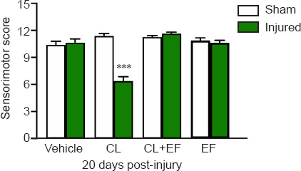
Norepinephrine plays an important role in motor functional recovery after a brain injury caused by ferrous chloride. Inhibition of norepinephrine release by clonidine is correlated with motor deficits after motor cortex injury. The aim of this study was to analyze the role of α2-adrenergic receptors in the restoration of motor deficits in recovering rats after brain damage. The rats were randomly assigned to the sham and injury groups and then treated with the following pharmacological agents at 3 hours before and 8 hours, 3 days, and 20 days after ferrous chloride-induced cortical injury: saline, clonidine, efaroxan (a selective antagonist of α2-adrenergic receptors) and clonidine + efaroxan. The sensorimotor score, the immunohistochemical staining for α2A-adrenergic receptors, and norepinephrine levels were evaluated. Eight hours post-injury, the sensorimotor score and norepinephrine levels in the locus coeruleus of the injured rats decreased, and these effects were maintained 3 days post-injury. However, 20 days later, clonidine administration diminished norepinephrine levels in the pons compared with the sham group. This effect was accompanied by sensorimotor deficits. These effects were blocked by efaroxan. In conclusion, an increase in α2-adrenergic receptor levels was observed after injury. Clonidine restores motor deficits in rats recovering from cortical injury, an effect that was prevented by efaroxan. The underlying mechanisms involve the stimulation of hypersensitive α2-adrenergic receptors and inhibition of norepinephrine activity in the locus coeruleus. The results of this study suggest that α2 receptor agonists might restore deficits or impede rehabilitation in patients with brain injury, and therefore pharmacological therapies need to be prescribed cautiously to these patients.
Keywords: alpha2-adrenoceptors; ambulatory behavior; clonidine; cortical injury; efaroxan; functional recovery; immunohistochemistry; motor deficit; norepinephrine; sensorimotor score.
Conflict of interest statement
None
Figures


Similar articles
-
Analysis of the receptor involved in the central hypotensive effect of rilmenidine and moxonidine.Naunyn Schmiedebergs Arch Pharmacol. 1999 Apr;359(4):262-71. doi: 10.1007/pl00005351. Naunyn Schmiedebergs Arch Pharmacol. 1999. PMID: 10344524
-
Neuronal norepinephrine responses of the rostral ventrolateral medulla and nucleus tractus solitarius neurons distinguish the I1- from the alpha2-receptor-mediated hypotension in conscious SHRs.J Cardiovasc Pharmacol. 2005 Jul;46(1):52-62. doi: 10.1097/01.fjc.0000162773.54915.52. J Cardiovasc Pharmacol. 2005. PMID: 15965355
-
The effects of the alpha2-adrenergic receptor agonists clonidine and rilmenidine, and antagonists yohimbine and efaroxan, on the spinal cholinergic receptor system in the rat.Basic Clin Pharmacol Toxicol. 2004 Apr;94(4):153-60. doi: 10.1111/j.1742-7843.2004.pto940401.x. Basic Clin Pharmacol Toxicol. 2004. PMID: 15078339
-
Noradrenergic modulation of XII motoneuron inspiratory activity does not involve alpha2-receptor inhibition of the Ih current or presynaptic glutamate release.J Appl Physiol (1985). 2005 Apr;98(4):1297-308. doi: 10.1152/japplphysiol.00977.2004. Epub 2004 Dec 3. J Appl Physiol (1985). 2005. PMID: 15579572
-
Norepinephrine release from spinal synaptosomes: auto-alpha2 -adrenergic receptor modulation.Anesthesiology. 2000 Jul;93(1):164-72. doi: 10.1097/00000542-200007000-00027. Anesthesiology. 2000. PMID: 10861160
References
-
- Ahmed-Farid OA, Haredy SA, Niazy RM, Linhardt RJ, Warda M. Dose-dependent neuroprotective effect of oriental phyto-derived glycyrrhizin on experimental neuroterminal norepinephrine depletion in a rat brain model. Chem Biol Interact. 2019;308:279–287. - PubMed
-
- Alcántara-Hernández R, Hernández-Méndez A. Adrenergic signaling molecular complexes. Gac Med Mex. 2018;154:223–235. - PubMed
-
- Apostolakis S, Kypraiou AM. Iron in neurodegenerative disorders:being in the wrong place at the wrong time? Rev Neurosci. 2017;28:893–911. - PubMed
-
- Aston-Jones G, Cohen JD. Adaptive gain and the role of the locus coeruleus–norepinephrine system in optimal performance. J Comp Neurol. 2005;493:99–110. - PubMed
LinkOut - more resources
Full Text Sources
