. 2022 Oct 18;11(20):e027593.
doi: 10.1161/JAHA.122.027593. Epub 2022 Oct 7.

Arteriovenous Fistulae in Chronic Kidney Disease and the Heart: Physiological, Histological, and Transcriptomic Characterization of a Novel Rat Model

Affiliations

Arteriovenous Fistulae in Chronic Kidney Disease and the Heart: Physiological, Histological, and Transcriptomic Characterization of a Novel Rat Model

Jef Van den Eynde et al. J Am Heart Assoc. .

Abstract

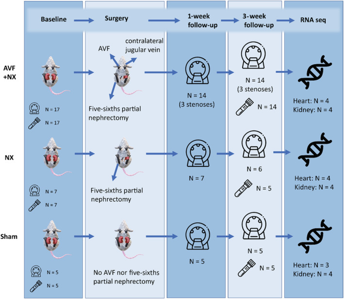
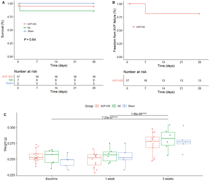
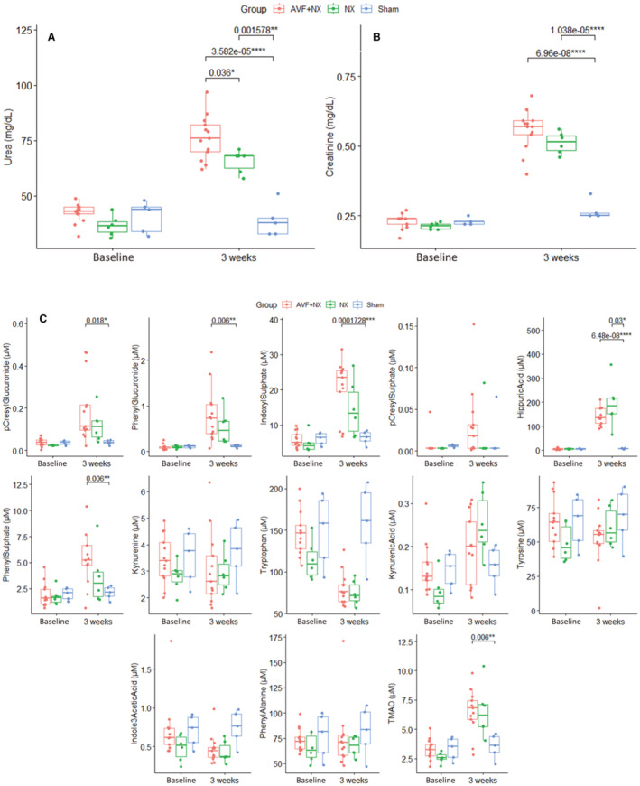
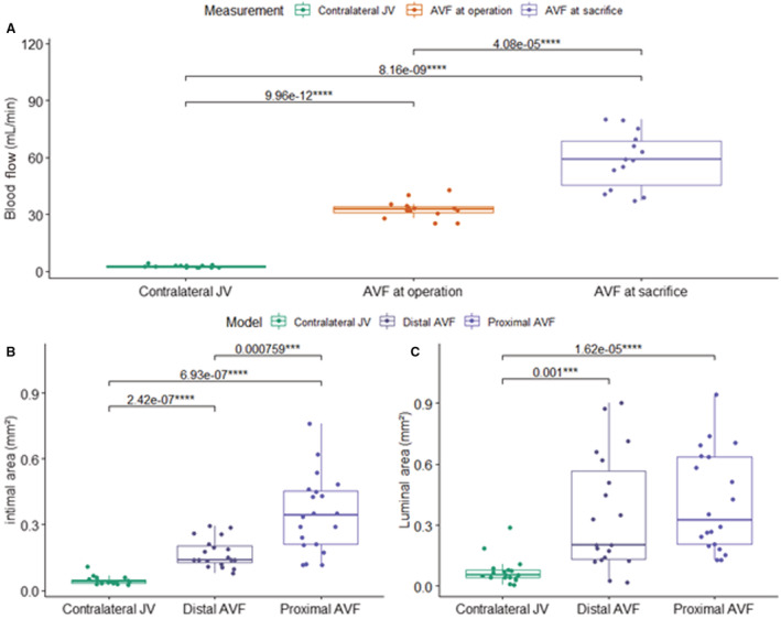
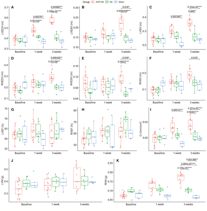
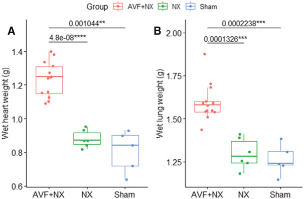
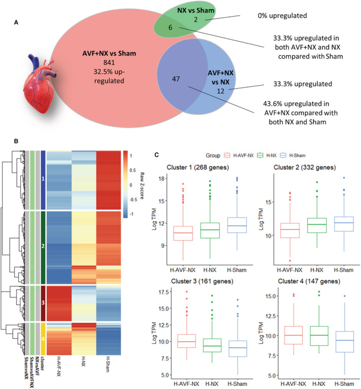
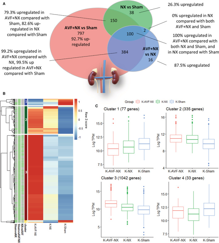
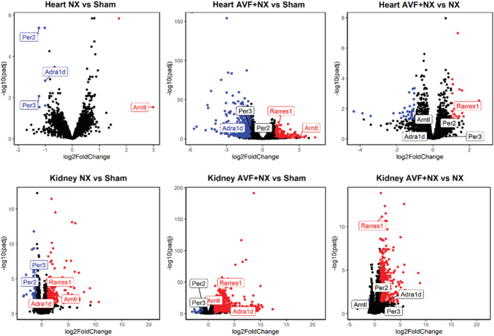
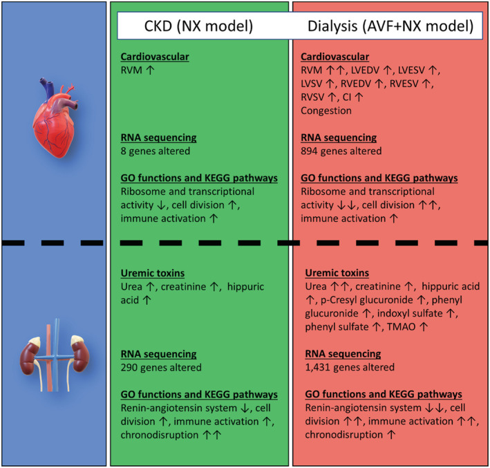
Background Arteriovenous fistulae (AVFs) are the gold standard for vascular access in those requiring hemodialysis but may put an extra hemodynamic stress on the cardiovascular system. The complex interactions between the heart, kidney, and AVFs remain incompletely understood. Methods and Results We characterized a novel rat model of five-sixths partial nephrectomy (NX) and AVFs. NX induced increases in urea, creatinine, and hippuric acid. The addition of an AVF (AVF+NX) further increased urea and a number of uremic toxins such as trimethylamine N-oxide and led to increases in cardiac index, left and right ventricular volumes, and right ventricular mass. Plasma levels of uremic toxins correlated well with ventricular morphology and function. Heart transcriptomes identified altered expression of 8 genes following NX and 894 genes following AVF+NX, whereas 290 and 1431 genes were altered in the kidney transcriptomes, respectively. Gene ontology and Kyoto Encyclopedia of Genes and Genomes analysis revealed gene expression changes related to cell division and immune activation in both organs, suppression of ribosomes and transcriptional activity in the heart, and altered renin-angiotensin signaling as well as chronodisruption in the kidney. All except the latter were worsened in AVF+NX compared with NX. Conclusions Inflammation and organ dysfunction in chronic kidney disease are exacerbated following AVF creation. Furthermore, our study provides important information for the discovery of novel biomarkers and therapeutic targets in the management of cardiorenal syndrome.

Keywords: animal model; arteriovenous fistula; cardiorenal syndrome; chronic kidney disease; heart failure.


Figures

Figure 1
Figure 1. Surgical procedures.
Microsurgical anastomosis of the cranial end of the right jugular vein to the side of the right carotid artery, as a model of brachial AVFs used for hemodialysis (A through F) and NX consisting of full removal of the right kidney and two‐thirds resection of the left kidney, as a model of CKD (G through I). AVF indicates arteriovenous fistula; CKD, chronic kidney disease; and NX, five‐sixths partial nephrectomy.
Figure 2
Figure 2. Study design.
AVF indicates arteriovenous fistula; and NX, five‐sixths partial nephrectomy.
Figure 3
Figure 3. Survival curves for overall survival (A) and freedom from AVF failure (B), and evolution of body weight during the course of the experiments (C).
AVF indicates arteriovenous fistula; and NX, five‐sixths partial nephrectomy. ****P≤0.0001.
Figure 4
Figure 4. Evaluation of CKD development from plasma assays of urea (A), creatinine (B), and a panel of uremic toxins and amino acids (C).
AVF indicates arteriovenous fistula; CKD, chronic kidney disease; NX, five‐sixths partial nephrectomy; and TMAO, trimethylamine N‐oxide. *P≤0.05; **P≤0.01; ***P≤0.001; ****P≤0.0001.
Figure 5
Figure 5. Blood flow measurements (A) and histological analysis of the AVF in the AVF+NX model at the time of euthanasia (B and C).
AVF indicates arteriovenous fistula; JV, jugular vein; and NX, five‐sixths partial nephrectomy. ***P≤0.001; ****P≤0.0001.
Figure 6
Figure 6. Magnetic resonance measurements of cardiac function and morphology.
(A) LVEDV, (B) LVESV, (C) LVSV, (D) RVEDV, (E) RVESV, (F) RVSV, (G) LVEF, (H) RVEF, (I) CI, (J) LVM, (K) RVM. AVF indicates arteriovenous fistula; CI, cardiac index; EDV, end‐diastolic volume; EF, ejection fraction; ESV, end‐systolic volume; LV, left ventricular; M, mass; NX, five‐sixths partial nephrectomy; RV, right ventricular; and SV, stroke volume. *P≤0.05; **P≤0.01; ***P≤0.001; ****P≤0.0001.
Figure 7
Figure 7. Postmortem measurements of “wet” heart (A) and lung weights (B).
AVF indicates arteriovenous fistula; and NX, five‐sixths partial nephrectomy. **P≤0.01; ***P≤0.001; ****P≤0.0001.
Figure 8
Figure 8. Genes altered in the heart of AVF+NX, NX, and Sham animals (n=1487, P<0.05 after adjustment for multiple comparisons, absolute log2 fold change >1).
(A) A total of 8 genes differed between NX and Sham, 894 genes differed between AVF+NX and Sham, and 59 genes differed between AVF+NX and NX. Percentages indicate the number of genes upregulated in the former of the 2 groups. (B) Heatmap representing mean transcripts per million (TPM) from all samples in the AVF+NX, NX, and Sham groups for genes differentially expressed between these groups. TPM was scaled using Z scores to allow for genes expressed at high and low abundance to be visualized on the same graph. For each gene, the color scale indicates high (red) or low (blue) expression relative to other groups. Green bars indicate significant differences between groups. Unsupervised hierarchical clustering identified 4 clusters with distinct patterns of expression across groups. (C) Box plots summarizing gene expression for all genes in each cluster in (B), based on the mean log TPM values for all samples in each group. AVF indicates arteriovenous fistula; and NX, five‐sixths partial nephrectomy.
Figure 9
Figure 9. Genes altered in the kidney of AVF+NX, NX, and Sham animals (n=1487, P<0.05 after adjustment for multiple comparisons, absolute log2 fold change >1).
A, A total of 290 genes differed between NX and Sham, 1431 genes differed between AVF+NX and Sham, and 502 genes differed between AVF+NX and NX. Percentages indicate the number of genes upregulated in the former of the 2 groups. B, Heatmap representing mean transcripts per million (TPM) from all samples in the AVF+NX, NX, and Sham groups for genes differentially expressed between these groups. TPM was scaled using Z scores to allow for genes expressed at high and low abundance to be visualized on the same graph. For each gene, the color scale indicates high (red) or low (blue) expression relative to other groups. Green bars indicate significant differences between groups. Unsupervised hierarchical clustering identified 4 clusters with distinct patterns of expression across groups. C, Box plots summarizing gene expression for all genes in each cluster in (B), based on the mean log TPM values for all samples in each group. AVF indicates arteriovenous fistula; and NX, five‐sixths partial nephrectomy.
Figure 10
Figure 10. Volcano plot for each of the comparisons between groups (AVF+NX, NX, and Sham) for heart and kidney tissues.
Each data point in the scatter plot represents a gene. The log2 fold change of each gene is represented on the x axis, and the log10 of its adjusted P value is on the y axis. Genes with an adjusted P value <0.05 and a log2 fold change >1 (upregulated genes) are indicated by red dots, while those with a log2 fold change <−1 (downregulated genes) are indicated by blue dots. AVF indicates arteriovenous fistula; and NX, five‐sixths partial nephrectomy.
Figure 11
Figure 11. Summary of the key study findings.
AVF indicates arteriovenous fistula; CI, cardiac index; CKD, chronic kidney disease; EDV, end‐diastolic volume; EF, ejection fraction; ESV, end‐systolic volume; GO, gene ontology; KEGG, Kyoto Encyclopedia of Genes and Genomes; LV, left ventricular; M, mass; NX, five‐sixths partial nephrectomy; RV, right ventricular; SV, stroke volume; and TMAO, trimethylamine N‐oxide.

Similar articles

Cited by

References

    1. Groenewegen A, Rutten FH, Mosterd A, Hoes AW. Epidemiology of heart failure. Eur J Heart Fail. 2020;22:1342–1356. doi: 10.1002/ejhf.1858 - DOI - PMC - PubMed
    1. Heidenreich PA, Albert NM, Allen LA, Bluemke DA, Butler J, Fonarow GC, Ikonomidis JS, Khavjou O, Konstam MA, Maddox TM, et al. Forecasting the impact of heart failure in the United States: a policy statement from the American Heart Association. Circ Heart Fail. 2013;6:606–619. doi: 10.1161/HHF.0b013e318291329a - DOI - PMC - PubMed
    1. Jones NR, Roalfe AK, Adoki I, Hobbs FDR, Taylor CJ. Survival of patients with chronic heart failure in the community: a systematic review and meta‐analysis. Eur J Heart Fail. 2019;21:1306–1325. doi: 10.1002/ejhf.1594 - DOI - PMC - PubMed
    1. Damman K, Valente MAE, Voors AA, O'Connor CM, Van Veldhuisen DJ, Hillege HL. Renal impairment, worsening renal function, and outcome in patients with heart failure: an updated meta‐analysis. Eur Heart J. 2014;35:455–469. doi: 10.1093/eurheartj/eht386 - DOI - PubMed
    1. Foley RN, Parfrey PS, Sarnak MJ. Clinical epidemiology of cardiovascular disease in chronic renal disease. Am J Kidney Dis. 1998;32:112–119. doi: 10.1053/ajkd.1998.v32.pm9820470 - DOI - PubMed

Publication types